
ligament pain
Ligament Pain in the Knee: How to Choose Between Self-Care, Evaluation, and Urgent Help
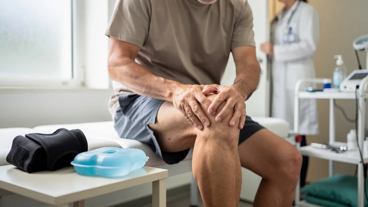
Start with the decision your symptoms can support
Possible ligament pain in your knee calls for a decision about what to do next, not a diagnosis made from a symptom list. If you are in Palo Alto or elsewhere in the Bay Area, start by sorting your symptoms into one of three paths: urgent medical evaluation, a timely knee assessment, or brief conservative care while you watch for change.
The details that matter most are practical. How did the pain begin? Could you bear weight afterward? Did the knee swell quickly, give way, lock, or lose movement? Did a pop occur with sudden weakness? Those answers help determine whether home measures are reasonable for a short period or whether you need qualified help sooner.
Use this sequence:
- Screen for warning signs. Loss of function, rapid swelling, marked instability, deformity, numbness, fever with redness and swelling, or severe injury-related pain should move you toward urgent evaluation.
- If no warning sign is present, reduce aggravating activity. Brief rest, gentle movement, and cautious comfort measures may fit a lower-risk recent injury.
- Arrange an evaluation when improvement stalls or function remains limited. A professional can examine the knee and decide whether imaging is useful.
- Discuss PRP, BMAC, or another non-surgical option only after the knee has been assessed. Symptoms alone cannot establish whether a regenerative treatment fits your injury.
This approach is especially useful when you want to keep hiking, cycling, playing sports, or managing daily activity without rushing into a treatment choice. It also prevents a common mistake: treating a knee that is unstable, blocked, or rapidly swelling as though it were only sore.
A symptom pattern can tell you which level of attention deserves consideration, but it cannot identify the injured ligament or determine whether you qualify for a particular treatment. Use the warning-sign section before trying self-care.
Put ligament-area knee pain in context
Knee injuries occur in sports and can also follow a minor accident, such as misjudging a step. That matters in an active area such as Palo Alto and the wider Bay Area, where running, court sports, cycling, hiking, and recreational training can all place different demands on the knee. A painful knee does not require a dramatic collision to deserve attention.
Knee pain also affects adults across age groups. A cited study examined knee-pain prevalence among adults age 18 and older, by age and sex, and reported that prevalence was high and had increased over time across age groups, with the largest increases among adults ages 25–44 and 45–64. This is context about knee pain overall, not a personal estimate of ligament injury or a measure of how common ligament pain is in Palo Alto.
You may notice a change in the character of pain after an injury. One patient-information source describes knee pain as often sharp at first and commonly becoming an ache within 10 days. That change alone does not show that the knee has healed, and it does not tell you whether the problem involves a ligament, cartilage, meniscus, muscle, or another structure. Pay attention to function as well as pain intensity.
A separate set of figures comes from cited school-athlete knee-injury data, so it should not be applied to all adults or treated as the frequency of ligament pain. In that study, knee injuries involved several structures, including the medial collateral ligament, or MCL, the ACL, the meniscus, the lateral collateral ligament, or LCL, and the posterior cruciate ligament, or PCL.
Those study figures describe the share of injuries involving each structure in that study population, and the categories can overlap. They do not predict which structure is affected in your knee. Use them to understand why an examination is more useful than guessing from the location of pain.
The useful question is not, “How common is my exact problem?” It is, “What changed, what can my knee do now, and is the pattern improving?” A twist during a weekend soccer match, a fall on a trail near Palo Alto, and gradually increasing pain after months of activity may all need different questions at an evaluation.
Read the pattern without naming the injury
A knee sprain occurs when fibers in a knee ligament overstretch or tear. Ligaments connect and support bones and help keep the knee stable while it bends and rotates. Sports injuries and falls commonly cause sprains, but an awkward landing, sudden twist, direct impact, excessive pressure, or a minor accident can also stretch a ligament too far.
That description explains the type of tissue involved without telling you which tissue is injured. Pain on the inside or outside of your knee may come from different structures, and pain near the joint does not prove that a ligament is the source.
Possible symptoms of a sprain or strain include:
- Pain, bruising, or swelling.
- Difficulty bending, straightening, or using the knee normally.
- A feeling that the joint is unstable.
- A sharp or deep pain after a specific movement.
- A pop or popping sensation at the time of injury.
- Buckling when you try to stand or put weight through the leg.
A cruciate ligament injury may cause a pop, buckling, and swelling without causing much pain at first. A collateral ligament injury can cause the knee to pop and buckle, followed by pain and swelling. Symptoms vary, so the absence of one feature does not rule out a meaningful injury.
Timing gives the pattern more context. Sudden pain during a particular activity points toward an acute structural injury, while pain that builds over days or weeks may fit overuse, tendinitis, bursitis, or early arthritis. Swelling that appears quickly and swelling that appears several hours later can also raise different clinical questions.
Write down what happened while the details are fresh:
- The movement or impact that started the pain.
- Whether you heard or felt a pop.
- How soon swelling appeared.
- Whether you could walk and bear weight.
- Whether the knee has buckled, caught, or locked.
- Whether you have had an earlier injury, surgery, or arthritis.
The injury mechanism can help point toward the structure involved, but it does not establish a diagnosis by itself. Do not test the knee repeatedly to see whether it gives way.
Use the injury mechanism to frame the evaluation
The movement that preceded your knee pain can help a clinician decide what to examine. A sudden stop, twist, pivot, rapid change of direction, hyperextension, or impact to the outside of the knee or lower leg can occur with ACL injuries. A direct impact to the front of the knee or a hard landing on a bent knee is a pattern associated with PCL injuries. A blow from the outside or inside, or an awkward twist, can affect a side ligament such as the MCL or LCL.
These are examination prompts, not labels for you to apply at home. The same movement can load more than one structure, and a gradual pain pattern may point away from a single traumatic ligament event. Tell the examiner exactly what your foot, knee, and body were doing when symptoms began.
A cited review states that ACL lesions are usually produced by non-contact movements and that female athletes had a 4 to 6 times higher risk than male athletes for this type of injury. That figure measures relative risk in the reviewed athletic context; it does not determine your individual risk or establish that your pain involves the ACL.
If you play soccer, basketball, football, tennis, skiing, or another sport with stopping and pivoting, include your training schedule and the exact movement that caused the problem. If you are an active adult who developed pain during hiking, lifting, or a daily movement, describe that setting too. The history helps the evaluation stay focused without turning a mechanism into a diagnosis.

Treat loss of function and rapid change as warning signals
Seek urgent medical evaluation rather than relying on routine self-care if you have any of these signs:
- You cannot bear weight through the leg.
- The knee gives way, feels unstable, or buckles under load.
- The knee develops marked swelling or substantial swelling immediately after the injury.
- You cannot fully bend or straighten the knee, or it feels blocked.
- You notice an obvious deformity.
- You have numbness or pins and needles below the injured knee.
- You have fever together with knee redness, pain, and swelling.
- You have severe pain associated with an injury.
- You heard or felt a pop followed by sudden weakness or loss of function.
These signs describe a need for prompt assessment, not a specific diagnosis. Inability to bear weight warrants evaluation even if pain later eases.
Swelling within the first hour may signal blood inside the joint and warrants prompt evaluation. Swelling that appears hours later is described as more consistent with a meniscal injury or mild ligament sprain, but it still deserves close monitoring. Locking or catching can raise concern for a meniscal tear or loose body, while buckling under load can be associated with an ACL injury, patellar instability, or significant collateral-ligament damage.
Do not wait for a home-care trial when the knee is changing quickly or losing function. Arrange qualified help and explain the timing of the swelling, the movement that caused it, and what your knee can no longer do.
A brace, ice, or a short period of reduced activity should not delay care when your knee is blocked, unstable, numb, substantially swollen, deformed, or unable to bear weight. Those measures may provide comfort, but they do not answer the urgent question of what has happened.
Use brief, measured care for a lower-risk recent injury
If you have none of the warning signs above and the injury is recent, use a short period of measured care rather than either pushing through pain or keeping the knee completely still. For the first 24 to 72 hours after an injury, rest the knee without long periods of complete inactivity. Gently move it for 30 seconds every hour while you are awake.
The goal is limited protection while you observe how the knee behaves. Movement should remain gentle and should not become a test of strength, stability, or pain tolerance.
Reduce the activity that aggravates your knee. Complete rest is not always necessary; switching to lower-impact activity may help maintain movement while the knee calms down. After 72 hours, use the leg more if symptoms are settling, but avoid sports or heavy lifting until you have less pain and good movement.
For an active adult in the Bay Area, that may mean replacing a run, game, steep hike, or strength session with ordinary movement that does not aggravate the knee. It does not mean forcing a painful range or repeatedly checking whether you can pivot. If ordinary movement is difficult, weight-bearing becomes harder, or the knee starts to give way, move from self-care to evaluation.
Set a clear checkpoint instead of leaving self-care open-ended. Ask whether pain, swelling, walking, bending, and straightening are improving. If the answer is no, if symptoms return whenever you resume normal activity, or if the knee remains limited, arrange a qualified evaluation.

Apply comfort measures while acknowledging variable guidance
Guidance on icing after a knee injury varies. One source describes 15 minutes every two hours during the first 24 to 72 hours. Another suggests 10 to 15 minutes every two to three hours for a recent injury or swelling. A separate source recommends an ice pack for 10 to 15 minutes every hour during the first day, followed by every three to four hours for two or three days or until the pain goes away.
Because icing schedules vary, follow the instructions from a qualified clinician who understands your injury. Whichever schedule you are given, place a damp towel or other cloth barrier between wrapped ice and your skin to help avoid an ice burn. Stop and seek advice if swelling or pain is severe or worsening.
A brace or compression sleeve may help with mild swelling or instability and may make activity more comfortable. Treat it as support, not as proof that the knee is stable. It should not replace an evaluation when symptoms persist, movement remains limited, or the knee gives way.
Do not use support to return immediately to cutting, jumping, heavy lifting, or a sport that reproduces the injury. A comfortable knee can still have a problem that needs examination.
Pain medicine can help you keep moving, but discuss medication with a doctor, especially if you take other medication. Your medical history and current medicines affect that decision.

Arrange a timely knee evaluation when improvement stalls
A knee that locks, catches, gives way, or swells soon after an injury also deserves more than an indefinite home-care trial. Bring the timing, mechanism, swelling pattern, and activity limits to the appointment.
Recovery does not follow one calendar. Recovery depends on the type and severity of the sprain, rehabilitation, and the sports involved. Milder Grade I and Grade II MCL or LCL sprains generally heal within 2 to 4 weeks, while other knee sprains may take 4 to 12 months. Those durations measure general recovery ranges for specified sprain categories, not a promise about your knee.
Use these ranges as context, not as a personal countdown. The practical checkpoint is whether function is returning: can you bear weight, walk, bend, and straighten the knee, and are pain and swelling settling rather than worsening? A pop followed by weakness, immediate swelling, buckling, or difficulty bearing weight changes the decision even when the injury is recent. Keep a brief record of the mechanism, when swelling began, movements that remain limited, and how the knee responds to lower-impact activity. Include earlier injuries, surgery, arthritis, braces, rehabilitation, or other care. This gives the clinician a clearer account of what changed and whether the pattern is new, recurring, or unresolved. It also supports a focused decision about continued conservative care, examination, imaging, or discussion of another treatment option.
Treatment decisions need individual context. A professional considers the injury history, stability, ability to bear weight, movement, severity when it can be established, associated findings, activity demands, and response to care. These details help determine whether continued conservative care, further assessment, or another treatment path should be discussed. Many knee ligament injuries do not require surgery, while more serious tears may require repair or reconstruction. Pain intensity or a symptom list alone cannot determine an individual treatment path. Previous knee problems can also change which causes are considered during evaluation.
Tell the clinician about earlier knee injuries, surgeries, arthritis, and the activities you want to return to. Previous knee problems can change which causes are considered during evaluation. If you have already tried rest, rehabilitation, a brace, or other care, describe what changed and what did not. That information helps separate a new event from a problem that needs a different assessment.
Know what the evaluation is designed to clarify
A qualified knee evaluation usually begins with your history: what movement or impact occurred, how quickly symptoms appeared, whether you heard a pop, and whether you could bear weight. The examination may check for swelling, pain, tenderness, warmth, bruising, movement, and the integrity of the knee’s structures.
Palpation maps tenderness to anatomical landmarks. Joint-line tenderness can point toward the meniscus, while tenderness at ligament insertion points can suggest collateral-ligament involvement. A qualified examiner may use a valgus stress test to assess the MCL; pain or laxity with inward force is a positive finding. A varus stress test assesses the LCL; pain or laxity with outward force is a positive finding. These maneuvers belong in a professional examination, not a self-test at home.
The knee relies on four main ligaments: the medial and lateral collateral ligaments at the sides and the cruciate ligaments within the joint. A history of a twist, impact, sudden stop, or fall may guide which structures receive closer attention, but the mechanism does not establish the diagnosis. The same symptom can occur with more than one knee problem, which is why the examiner considers movement, stability, swelling, and tenderness together. Share whether symptoms began suddenly or gradually, whether swelling appeared immediately or hours later, and whether the knee has buckled, caught, or locked. Also mention any earlier injury, surgery, arthritis, or care already tried. Those details help the clinician decide which examination findings matter and whether an X-ray or MRI would answer a specific question.
Imaging answers different questions. An X-ray may look for a fracture, degenerative joint disease, arthritis, bone spurs, or fluid around the joint. An MRI is particularly useful for soft-tissue injuries involving ligaments, tendons, cartilage, or muscles. The choice depends on the history and examination rather than on the word “ligament” alone.
If you already have an X-ray, MRI, or earlier treatment report, bring it if available. Imaging is interpreted alongside your symptoms and physical findings; it is not a substitute for examining how the knee moves and functions.
A clinician may describe a collateral sprain by grade. Grade 1 means the ligament is stretched but remains intact and stable. Grade 2 involves partial tearing with some joint laxity. Grade 3 is a complete rupture.

Keep treatment decisions after the assessment
Many knee ligament injuries do not require surgery. Mild injuries can sometimes be treated with rest and possibly doctor-guided physical therapy, while treatment for other injuries may include medication, a brace, repair, or reconstruction. The relevant choice depends on the injury, severity, stability, function, associated findings, activity demands, and response to care.
Most Grade 1 and Grade 2 collateral ligament injuries are described as responding well to conservative care. Grade 3 tears, particularly ACL tears, often require surgical reconstruction depending on age, activity level, and associated injuries. These are general treatment patterns, not a decision about your knee. A professional must connect the grade and examination findings to your goals and options.
Your desired activity matters, but wanting to return to hiking, tennis, skiing, or competitive sport does not by itself determine the treatment. Some injuries can be managed without surgery; some require a different level of care to restore function. Do not choose a regenerative injection simply because you want to avoid surgery, and do not assume that surgery is necessary because the knee hurts.
The next decision should follow the assessment: continue guided conservative care, obtain additional imaging or specialist input, discuss a non-surgical option, or address an injury that needs conventional treatment. Urgent warning signs take priority over all of these discussions.
When a clinician discusses options, compare them using the same questions rather than comparing treatment names alone. First ask what problem the plan is meant to address: pain, loss of motion, instability, or a structural finding. The answer should connect to the history and examination, not only to where the knee hurts. Next ask what makes the option appropriate now: the established or suspected severity, stability, associated findings, activity demands, and response to the care already tried.
Then clarify the decision’s time horizon. Is the proposed step intended to support continued conservative care, answer an unresolved question, restore function, or address a problem that may need repair or reconstruction? Those are different purposes. A treatment discussion is easier to understand when the purpose and the next checkpoint are explicit, rather than when the option is presented as a general solution for knee pain.
Your activity goal belongs in the conversation, but it is one factor among several. A person hoping to return to Bay Area hiking, cycling, court sports, or another active routine can ask how the findings affect that goal and what limitations should remain while the knee is being evaluated or treated. That question does not turn a desired activity into a treatment indication.
If PRP, BMAC, or another regenerative option is discussed, ask which evaluation findings support discussing it, which findings would make another approach more appropriate, and what remains uncertain. The fact that a treatment is available does not establish suitability for a particular ligament-area problem. Keep urgent or necessary conventional care ahead of a non-urgent treatment preference.
A sound next-step conversation should leave you knowing what is being considered, why it is being considered, what alternatives remain, and what change would prompt reassessment. If those points are unclear, ask for the reasoning in plain language before choosing among options. The goal is not to select a treatment from a list; it is to match the next decision to the knee’s documented function, examination, and clinical question.
Discuss regenerative options with realistic expectations
At RegenCore Method, we provide research-backed, non-surgical treatments including PRP, bone marrow concentrate (BMAC), stem cell therapy, shockwave, EMTT, and spinal disc care. We treat conditions affecting tendons, ligaments, cartilage, muscles, and joints due to osteoarthritis and injury. We commonly use PRP for conditions including knee osteoarthritis, patellar and Achilles tendinopathy, and hip and gluteal tendon pain.
For possible ligament pain, that information describes options available for discussion; it does not establish that PRP, BMAC, or another regenerative treatment is suitable for your knee. Suitability depends on an individualized evaluation of the injury, stability, function, associated conditions, and prior care. Symptoms alone cannot determine the treatment plan, and a regenerative option should not delay urgent or necessary conventional care.
Cost information also requires a clear boundary. A cited pricing source reports that single-joint PRP procedures generally fall in the $500 to $1,500 range per injection. The same source states that PRP at Regenerative Joint Clinics in Riverdale, Georgia, was $500 per injection, with total cost depending mainly on the number of injections recommended.
Those figures measure pricing reported by a separate clinic in Riverdale, Georgia, not RegenCore Method pricing, insurance coverage, or a Palo Alto estimate. Do not use them to predict your cost. The number of injections, the treatment under discussion, and the clinical plan must be addressed in the appropriate evaluation and financial conversation.
Before discussing PRP or BMAC, bring focused questions:
- What structure or structures appear involved, and what remains uncertain?
- What did the examination show about stability and movement?
- Is imaging needed to answer a specific question?
- What conventional and non-surgical options fit the findings?
- What would make urgent or surgical care more appropriate?
- How would progress be assessed if you choose conservative or regenerative care?
This keeps the discussion tied to your knee rather than to a treatment name.
Choose the next conversation in Palo Alto or the Bay Area
Possible ligament pain is best handled in order: screen for loss of function and rapid swelling, use brief measured care only when warning signs are absent, arrange an evaluation when improvement stalls, and discuss treatment options after the examination. If you are seeking a knee-pain assessment in Palo Alto or the Bay Area, bring the injury timing, movement or impact, swelling pattern, walking limits, episodes of buckling or locking, prior imaging, and treatments you have already tried.
A qualified professional must decide individual suitability for evaluation, imaging, rehabilitation, surgery, PRP, BMAC, or another treatment. If you have an urgent warning sign, seek urgent medical attention rather than waiting for a non-urgent treatment discussion.
A useful appointment starts with a clear account of what changed and what your knee can do now. That information gives the conversation a practical starting point and helps you choose between continued monitoring, a timely assessment, or urgent care.
Questions to ask before arranging a knee evaluation
How common is ligament pain in the knee, and who is most affected?
Knee pain is common across adult age groups, while ligament injuries are often reported after sports, falls, twists, impacts, or awkward landings. Sport-specific injury figures should not be applied to every adult or used to predict an individual diagnosis.
Can knee ligament pain happen without a major injury?
Yes. A ligament-area problem can follow a minor accident, misjudged step, awkward landing, or sudden twist, and age-related wear may make a small event trigger a pain flare. Gradual pain may instead raise questions about overuse, tendinitis, bursitis, arthritis, or another structure, so the pattern alone does not identify the cause.
When should I seek urgent help for possible ligament pain?
Seek urgent medical evaluation if you cannot bear weight, the knee gives way or feels unstable, swelling is marked or develops immediately, the joint is blocked or cannot move normally, there is deformity, numbness below the knee, fever with redness and swelling, or severe injury-related pain. A pop followed by sudden weakness or loss of function also warrants prompt assessment.
Could PRP or BMAC be appropriate for ligament pain?
Possibly, but symptoms alone cannot establish suitability. A qualified evaluation should first consider the injury mechanism, stability, movement, associated findings, prior care, and whether imaging is needed. PRP or BMAC should not delay urgent care, necessary conventional treatment, or treatment for an injury that may require a different approach.
Next step
Have a question for RegenCore?
Concierge regenerative & orthobiologic orthopedics in San Francisco and Palo Alto. Research-backed, non-surgical treatment — PRP, bone marrow concentrate (BMAC), stem cell therapy, shockwave, EMTT and spinal disc care — to help the body heal itself.
Get in touch