
articular cartilage pain
Articular Cartilage Pain: How to Choose a Safe Next Step for Knee Symptoms
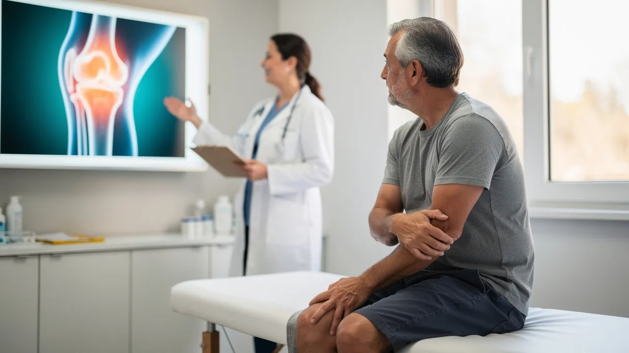
Begin with what cartilage-related knee symptoms can—and cannot—tell you
Articular cartilage covers the ends of bones in a joint, cushions the joint, and helps the bones glide smoothly during movement. When that tissue is injured by trauma or affected by progressive degeneration, your knee may develop pain, swelling, stiffness, limited motion, or a locking or catching sensation. Pain may wax and wane rather than follow a steady pattern.
Cartilage-related symptoms can make walking, running, or other activities difficult, and recurring swelling can be a frequent warning sign. They still do not identify the exact source of your knee pain. Ligaments, the meniscus, alignment, bone, and other joint tissues can produce overlapping symptoms, so treat the pattern as a reason to sort the next step—not as a diagnosis.
Knee cartilage surgery has a long recovery window when it becomes appropriate. Recovery can take up to one year, and recovery can take up to 18 months when the patella is involved. These timeframes describe cartilage procedures, not a prediction about your knee or a reason to assume surgery is necessary. An evaluation should establish what structure is involved and which choices are relevant before you compare treatment paths.
That fact explains why a persistent symptom pattern deserves attention, but it does not tell you whether your pain comes from cartilage, how extensive any injury may be, or which treatment would be appropriate. A painful knee after a hike, a twist, or a period of increasing stiffness still needs its history and examination interpreted together.
Put prevalence and risk into useful context
Articular cartilage injuries are common, especially among physically active people, and they occur most often in the knee. Osteoarthritis is the most common form of arthritis and affects approximately 90 million adults in the United States, or 36.8% of the U.S. adult population. Osteoarthritis primarily affects articular cartilage in load-bearing joints such as the knee, hip, and shoulder.
That context applies to active adults as well as people whose symptoms are connected with age-related wear. It does not tell you whether cartilage is the source of your pain.
Knee osteoarthritis risk is influenced by age, weight, prior joint trauma, and repetitive movements such as squatting and kneeling. Osteoarthritis affects people of all ages. A review reports that previous knee trauma increases the risk of knee osteoarthritis 3.86 times, while muscle weakness, joint laxity, repetitive use, overweight and obesity, and other factors also play roles.
A clinical overview reports that healthy people younger than 40 with minor articular cartilage injuries generally have the best outcomes and often return to pre-injury activities. The same overview describes cartilage restoration or repair procedures as intended for active people under age 50, but it also states that not everyone is suitable. Those age ranges are source-specific context, not a candidacy rule for you. Use your injury history, current function, examination, and relevant imaging to make the decision.
Common knee cartilage problems can affect very different people and follow different patterns. A physically active adult may notice symptoms after a direct blow, twist, repeated loading, or a change in training. Another person may notice gradually increasing stiffness during stairs, walking, or daily tasks. Age and activity can shape the context, but neither factor identifies the painful structure.
Use prevalence to understand why evaluation questions arise, not to label your symptoms. A history of knee trauma, repetitive kneeling, or increasing difficulty with ordinary activity makes the timeline worth documenting. It does not by itself establish osteoarthritis or a cartilage injury.
Use function and change over time as the first sorting questions
Knee function and symptom change over time are useful sorting questions when you cannot yet identify the cause. Osteoarthritis can cause pain and stiffness that worsen over time and make daily activities difficult. Knee arthritis may also involve swelling, pain after sitting or resting, flares with vigorous activity, creaking or grinding, locking or sticking, weakness, or buckling.
Knee pain should not automatically be attributed to osteoarthritis. Some people with osteoarthritis have no symptoms, and radiographic examination may not identify it because of limited sensitivity. Persistent pain, recurring swelling, loss of motion, locking, giving way, or difficulty walking or exercising makes an evaluation more useful than continued guesswork.
A new knee problem deserves professional attention if the knee swells immediately after a twisting injury, locks, or gives way. Contact a clinician if knee pain continues after 3 days of home treatment. These thresholds do not establish a diagnosis; they mark a change from watching a lower-risk pattern to finding out why symptoms are persisting or affecting function.
Before an appointment, note when the symptoms began, whether a fall or twist preceded them, where the pain is located, what movements make it worse, and whether swelling or mechanical symptoms occur. That information can help the evaluation focus on the questions that matter.
You can use four questions to decide whether brief monitoring still makes sense:
- Is the knee improving, unchanged, or worsening?
- Can you walk, bend, and straighten it as usual?
- Did the symptoms follow a fall, twist, or direct injury?
- Are swelling, locking, buckling, or repeated activity-related flares appearing?
Improvement and preserved function support a measured first response when no warning sign is present. Persistence, worsening, loss of function, or mechanical symptoms move the decision toward an evaluation. The questions sort urgency; they do not identify the exact diagnosis.
Make a measured first move for a new, lower-risk knee problem
A new, lower-risk knee problem calls for relative rest rather than complete inactivity during the first 24 to 48 hours. For that initial period, use the following measures:
- Rest from activities that cause pain, and avoid putting weight on the knee when weight-bearing is painful.
- Move the knee gently for 10 to 20 seconds every hour while you are awake instead of keeping it completely still.
- Apply ice for up to 15 minutes at a time with a towel barrier. For acute knee pain, apply ice every hour initially for up to 15 minutes; after the first day, apply it at least 4 times per day.
- Keep the knee raised as much as possible when swelling is present.
- Consider an elastic bandage or sleeve for support and possible swelling reduction.
These measures are for short-term symptom care. They do not show that cartilage is injured or repair damaged cartilage.
After 48 hours, a new knee problem may call for gradually using the leg more, provided discomfort is lower and movement is improving. Avoid sports or heavy lifting until you have less discomfort and good movement. A gradual return is different from testing the knee with a run, jump, deep squat, or heavy lift to see whether the problem has disappeared.
Stop treating the situation as routine self-care if pain remains severe, function is declining, the knee locks or gives way, or a new warning sign appears. The next step then becomes qualified assessment rather than adding more home measures.
Short-term care also has clear boundaries. Do not keep the knee completely still for long periods, leave ice on longer than the advised interval, or use a painful activity as a test of whether the knee is ready. A towel barrier matters when you apply ice, and support from an elastic sleeve or bandage should remain part of comfort care rather than a reason to resume heavy loading.
If you cannot bear weight, the knee is misshapen, or severe pain continues even without weight-bearing, move directly to prompt medical evaluation instead of extending home care.

Reduce avoidable aggravation while symptoms settle
Knee-friendly activity modification may help reduce aggravation while symptoms settle. Warm up before exercising, cool down afterward, and stretch your quadriceps and hamstrings. Reduce or modify painful exercise, avoid running down hills, and consider bicycling or swimming instead of running. If you run, choose a smooth, softer surface such as a track rather than cement or pavement, and use shoes that fit well, are well made, and have good cushioning.
Use pain, swelling, and function as reasons to reduce the activity further or arrange an evaluation.
Excess weight changes knee loading during certain movements. MedlinePlus reports that every pound, or 0.5 kilogram, of excess weight puts about 5 extra pounds, or 2.25 kilograms, of pressure on the kneecap when you go up and down stairs, with even greater pressure during jumping. That figure describes kneecap pressure in the specified activities; it is not a measure of your individual pain or cartilage damage.
After a fall or acute twisting knee injury, ultrasound is not routinely used as the initial imaging study in adults or children 5 years of age or older who have focal tenderness, an effusion, or inability to bear weight. Imaging choices depend on the injury pattern and examination, so do not choose a test simply because it is available.
The practical goal of activity modification is to change the movements that provoke symptoms while preserving comfortable movement. For example, replacing a painful run with bicycling or swimming avoids continuing the same painful activity. Avoiding downhill running and choosing a smoother surface also changes the demand placed on the knee.
These options are not a substitute for assessment when your walking, stairs, work, hiking, or exercise remain limited. A lower-impact activity may be reasonable to discuss or try within your comfort, but increasing symptoms, swelling, or loss of motion should change the plan.
Compare care pathways by what they are meant to accomplish
Knee care pathways answer different questions. Short-term self-care asks whether a new, lower-risk pattern settles with measured activity changes. Rehabilitation and other nonsurgical care focus on symptoms, strength, and mobility. A procedure discussion asks whether a specific intervention is reasonable after the knee problem has been assessed.
| Pathway | What it is meant to do | Boundary | Your next action |
|---|---|---|---|
| Short-term self-care and activity modification | Settle a new, lower-risk pattern and avoid painful loading | It cannot identify the cause or repair damaged cartilage | Use brief, measured care and track pain, swelling, motion, and function |
| Rehabilitation or other nonsurgical care | Relieve symptoms and improve strength or mobility; nonsurgical care may help prevent or slow further degeneration | It may improve symptoms without repairing damaged articular cartilage | Discuss a rehabilitation plan when symptoms persist or limit activity |
| PRP or BMAC discussion | Consider whether a procedure fits a specific diagnosis, goal, and alternative | Neither option is a default answer for every cartilage condition | Discuss the procedure only after an evaluation has clarified the knee problem |
Choose the next category by the question you need answered:
- If the problem is new, lower-risk, and improving, brief self-care can help you observe the trend without forcing activity.
- If pain, stiffness, swelling, or reduced movement persists, an evaluation can clarify whether rehabilitation or another symptom-focused plan fits.
- If you are considering PRP, BMAC, or another procedure, first establish what condition is being treated and what the procedure is intended to accomplish.
Each branch has a boundary. Self-care should not continue through warning signs or declining function, rehabilitation should not be treated as proof of a cartilage diagnosis, and a procedure should not be selected solely because symptoms have lasted a long time.
Treat PRP and BMAC as questions for an assessed knee, not default answers
A knee osteoarthritis management summary and RegenCore Method do not characterize PRP in the same way. A knee osteoarthritis management summary reports that PRP has very limited scientific support for helping with knee osteoarthritis. We offer PRP as a treatment that commonly helps knee osteoarthritis.
The useful conclusion is narrower: PRP belongs in a diagnosis-specific conversation, not in a default treatment sequence. Before discussing it, connect the symptoms with the examination, relevant imaging, the duration of the problem, your activity goals, and other available care.
BMAC is another procedure that should be considered only in the context of an assessed knee. We offer BMAC as an option that commonly helps cartilage injury in select candidates and moderate to advanced knee osteoarthritis. Our cell therapy information also includes cartilage injury in the right candidate among the situations it may help.
The phrases “select candidates” and “right candidate” set a real boundary. They do not establish that you qualify, that cartilage is the source of your symptoms, or that BMAC will avoid surgery. Bring those questions to a qualified evaluation, where the diagnosis, treatment alternatives, risks, and goals can be considered together.
Before a PRP or BMAC discussion, ask what condition the procedure is intended to address, what evidence applies to that condition, and what alternatives remain available. Also ask what result would count as meaningful for your goals, what uncertainty remains, and how the plan would change if the examination or imaging points to another source of pain.
Those questions keep a treatment conversation tied to your knee rather than to a general category of regenerative therapy. They also preserve the distinction between what a practice offers, what an external clinical summary reports, and what can be concluded about your individual situation.
Use cost ranges as orientation, not as a treatment quote
Stem cell therapy for knee pain has no single universal price. One published price guide reports a range from $3,000 to over $50,000, depending on treatment complexity, cell source, and geographic location. The same guide says insurance rarely covers regenerative treatments. That is a reported market range from one source, not a price for your evaluation, PRP, BMAC, or any particular treatment plan.
The guide reports $3,000 to $10,000 for localized treatment of simple joint pain and up to $20,000 to $50,000 for comprehensive multi-joint or systemic conditions. Those categories are not interchangeable with a quote for a single knee. Physician experience and condition complexity are also identified as pricing factors.
A useful cost conversation separates the treatment from the items surrounding it. Ask what the quoted amount covers, whether the estimate applies to one knee or more than one area, and which parts of evaluation, imaging, procedure, follow-up, or additional care are included. Ask how the estimate changes if the diagnosis or treatment plan changes.
Cost should be considered alongside the question the treatment is meant to answer. A low headline figure may describe a different procedure or scope of care than a higher figure, while a higher figure does not establish that an option is appropriate or effective for your condition.
The reported price ranges should be read as orientation rather than a local promise. The lower $3,000 to $10,000 range describes localized treatment for simple joint pain in that guide, while the $20,000 to $50,000 range describes comprehensive multi-joint or systemic conditions. A single knee with a specific diagnosis may not fit either category.
Cell source, treatment complexity, geographic location, and physician experience can all change the amount described by the guide. Insurance coverage is also not a safe assumption for regenerative treatment. Separate the cost question from the suitability question: knowing a price does not establish that the treatment addresses your pain.
Let the evaluation answer the cartilage question
A knee evaluation may connect your symptom history with an examination of motion, joint-line pain, swelling, fluid in the knee, alignment, and possible ligament or meniscal injury. Cartilage injuries can be difficult to diagnose. MRI or arthroscopy may be necessary, while X-rays are usually used to rule out other abnormalities rather than directly identify every cartilage problem.
Knee radiograph usefulness depends on the view. A review reports that standing flexed-knee images reliably showed tibiofemoral joint-space width and bone changes, while non-weight-bearing and weight-bearing images in extension had limited value for assessing disease status. The same review reports that skyline views were better than lateral views for detecting osteoarthritis-related changes in the patellofemoral joint. Existing imaging can be useful, but an image should be interpreted with your history and examination rather than treated as the whole diagnosis.
A sports-medicine source states that articular cartilage defects smaller than 2 cm have the best prognosis and treatment options. That statement concerns the defect size described by that source; it does not mean that a defect of any size determines your treatment or outcome. The location, depth, symptoms, other injuries, and overall knee condition still matter.
After a fall or acute twisting knee injury in an adult or child 5 years of age or older with focal tenderness, an effusion, or inability to bear weight, CT, MR arthrography, and bone scan with SPECT or SPECT/CT are not routinely used as the initial imaging studies. A clinician can decide whether another study is indicated after considering the mechanism, examination, and functional loss.
Prepare for an evaluation by bringing a short symptom timeline and any existing imaging reports or images that are available to you. Include:
- The date and mechanism of onset, including a fall, twist, direct blow, or gradual change.
- The activities that increase symptoms and the activities you can still do comfortably.
- Changes in swelling, motion, locking, catching, buckling, or walking tolerance.
- Previous rest, rehabilitation, injections, or other treatment and what changed afterward.
- Your practical goal, such as walking, stairs, hiking, cycling, or returning to a particular activity.
This information does not replace an examination. It helps connect the history, physical findings, and imaging to the decision rather than letting a scan or a symptom label determine the whole conversation.

Know which changes should move you out of routine self-care
Prompt medical evaluation is appropriate when knee symptoms include any of the following:
- You cannot bear weight, or you have severe pain even when you are not bearing weight.
- Your knee buckles, clicks, or locks; is deformed or misshapen; or cannot flex or fully straighten.
- Your knee has fever, redness or warmth around it, or a large amount of swelling.
- Your calf below the sore knee has pain, swelling, numbness, tingling, or bluish discoloration.
These changes can signal a problem that should not be sorted out through routine home care alone. Contact qualified medical help promptly rather than testing the knee with exercise or waiting for a procedure discussion to answer an urgent question.
A knee that swells immediately after a twisting injury, locks, or gives way also warrants prompt professional attention. Knee pain that remains after 3 days of home treatment is another reason to contact a clinician. The practical boundary is function and change: inability to use the leg normally, rapidly increasing symptoms, or concerning whole-body or calf signs moves you away from routine self-care.

Choose an evaluation conversation in Palo Alto or the Bay Area
A persistent or activity-limiting knee problem deserves a conversation that starts with the symptom pattern and asks what remains uncertain. We offer stem cell therapy for knee pain and other conditions, but an evaluation is needed before discussing whether it is relevant to your situation.
Those offerings do not make you a candidate or establish that a procedure will produce a particular result. A qualified clinician decides individual suitability after considering your history, examination, relevant imaging, diagnosis, goals, alternatives, and the limits of the available evidence. For a conversation in Palo Alto or the Bay Area, bring your symptom timeline, injury details, prior treatment response, and imaging reports if you have them.
The right next step may be measured self-care, rehabilitation, further evaluation, or a discussion of PRP, BMAC, or another option. The decision should follow the question your knee still needs answered, not a promise about cartilage repair or avoiding replacement.
Common questions about knee cartilage pain, evaluation, and treatment options
What should I do if knee pain comes with swelling, locking, or trouble bearing weight?
Seek prompt medical evaluation if you cannot bear weight, have severe pain, a deformed knee, locking or buckling, inability to flex or fully straighten it, fever with redness or warmth, major swelling, or calf pain, swelling, numbness, tingling, or bluish discoloration. Immediate swelling after a twisting injury also warrants professional attention. Avoid testing the knee with exercise or heavy lifting while waiting for guidance.
Can an MRI confirm that articular cartilage pain is causing my knee symptoms?
Not by itself. Knee pain can come from cartilage, the meniscus, ligaments, alignment, bone, or other tissues. A qualified evaluation combines your symptom and injury history with examination of motion, swelling, joint-line pain, alignment, and possible associated injuries. MRI or, in some cases, arthroscopy may be needed, while X-rays are often used to assess other abnormalities. An image should be interpreted with the examination rather than treated as a diagnosis on its own.
What is the evidence for PRP or BMAC for knee cartilage problems?
The evidence and clinical positions are not uniform. One selected knee osteoarthritis summary reports very limited scientific support for PRP, while RegenCore Method describes PRP as commonly helpful for knee osteoarthritis and BMAC as an option for cartilage injury in select candidates and moderate to advanced knee osteoarthritis. Neither treatment is appropriate for every cartilage condition, and neither should be selected without clarifying the diagnosis, alternatives, risks, goals, and remaining uncertainty.
What might regenerative treatment for knee pain cost?
One published price guide reports stem cell therapy for knee pain ranging from $3,000 to over $50,000. It reports $3,000 to $10,000 for localized treatment of simple joint pain and up to $20,000 to $50,000 for comprehensive multi-joint or systemic conditions. Treatment complexity, cell source, geographic location, and physician experience can change the amount, and the guide says insurance rarely covers regenerative treatment. These figures are not a quote for a particular knee evaluation, PRP, BMAC, or treatment plan.
How common is articular cartilage pain, and who is most often affected?
Articular cartilage injuries are common, especially among physically active people, and occur most often in the knee. Osteoarthritis is the most common form of arthritis, affecting approximately 90 million adults, or 36.8% of the U.S. adult population. Risk is influenced by factors such as age, weight, prior knee trauma, repetitive use, muscle weakness, and joint laxity, but people of all ages can be affected. Prevalence and risk factors cannot establish that cartilage is the source of an individual’s pain.
Next step
Have a question for RegenCore?
Concierge regenerative & orthobiologic orthopedics in San Francisco and Palo Alto. Research-backed, non-surgical treatment — PRP, bone marrow concentrate (BMAC), stem cell therapy, shockwave, EMTT and spinal disc care — to help the body heal itself.
Get in touch