
flex knee
Flex Knee: What Knee Flexion Means and When Movement Changes Need Attention
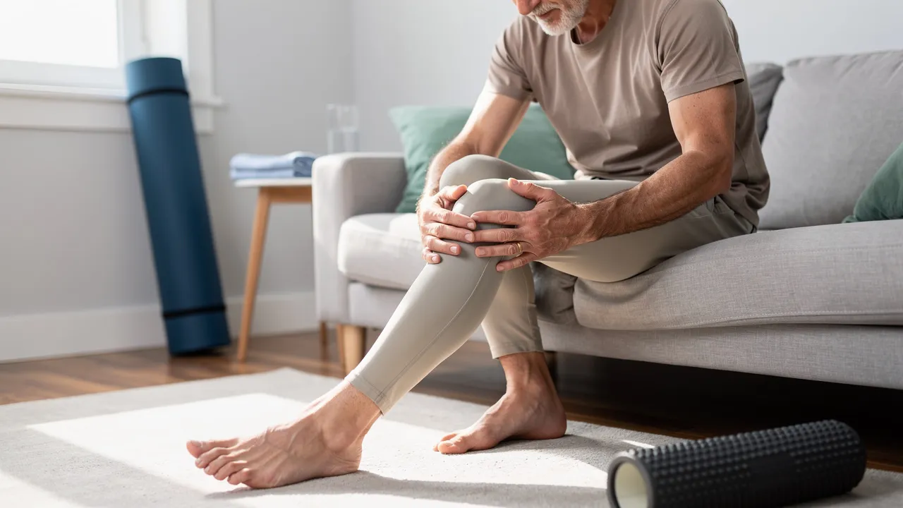
Start with what “flex knee” means
Knee flexion means bending your knee, while knee extension means straightening it. When you sit down, climb stairs, squat, or bring your heel toward your body, your knee is using flexion. When you stand from a chair or straighten your leg, your knee is using extension. Both movements are part of knee range of motion and support functional and sport-specific activities.
A clinical reference describes a normal knee range of motion that includes extension from 0 to −10° and flexion to 135°. That reference is a general clinical comparison, not a personal target or a diagnosis. Clinicians assess knee flexion and extension with active movement first and passive movement afterward, along with strength testing; the cited method grades strength from 0 to 5.
The phrase “flex knee” describes a movement rather than explaining why the movement feels painful, stiff, or limited. You can use the movement as a starting point: notice whether bending is different from straightening, whether one knee moves differently from the other, and whether symptoms change with walking, stairs, sitting, or activity. Those observations help frame the next question without turning one movement into an answer.
Active knee motion is the movement you produce yourself, while passive knee motion is assessed when a clinician moves the joint for you. The difference can give a clinician more context about pain, stiffness, strength, and the amount of movement available during an examination. You do not need to force either type of motion at home to make the comparison useful; describe what happens during ordinary movements and leave formal testing to a qualified professional.
Use a change in motion as a signal, not a verdict
Reduced knee flexion can affect gait, limit functional squatting, and make stairs and sitting difficult. You may notice the change as a shorter bend, a need to shift your weight, or difficulty completing a familiar movement. The practical meaning is functional: a change that interferes with ordinary activity deserves attention even when you cannot identify its cause.
A study comparing symptomatic and asymptomatic knees found that symptomatic knees had flexion from 45° to 130°, with a median of 105°, while asymptomatic knees ranged from 95° to 140°, with a median of 125°. The study group therefore showed overlap as well as difference; a range from a study cannot tell you what your own knee should measure.
A side-to-side change in knee flexion can be a useful signal to discuss, but it is not a personal cutoff. In one study of 110 patients comparing symptomatic and asymptomatic knees, 106 of 110 patients, or 96.4%, had at least a 10° reduction in flexion, while 88 of 110 patients, or 80%, had at least a 15° reduction. Your decision should depend on the movement change together with pain, swelling, function, injury history, and how the pattern develops.
A change in knee motion becomes easier to explain when you connect it to a specific task. Note whether you can sit down, rise from a chair, climb stairs, squat, walk, or participate in your usual activity in the same way as before. Difficulty with a task does not identify the cause, but it tells you how the movement change affects function and gives a clinician a clearer starting point.

Read the pattern around the movement
Limited or painful knee flexion can have several causes, including muscle tightness or imbalance, ligament or meniscus injuries, swelling or inflammation, osteoarthritis, an acute injury, a mechanical block, or stiffness after an earlier injury or surgery. Traumatic injuries such as ligament sprains, meniscal tears, or fractures can cause pain, swelling, and instability that reduce knee movement. These possibilities explain why movement alone cannot identify the cause.
The timing of knee symptoms gives you a useful observation point. Sudden onset during a specific activity points toward an acute structural injury, while gradual onset over days or weeks suggests overuse, tendinitis, bursitis, or early arthritis. In physically active people, changes in training intensity, duration, frequency, environment, or activity mode are frequent contributors to anterior knee pain. Anterior knee pain may increase with running, squatting, stairs, or prolonged sitting, and it often worsens when you continue the activity that provokes it.
Movement-related symptoms should be read as decision cues, not as a diagnosis. Swelling that appears hours after an injury is more consistent with a meniscal injury or mild ligament sprain. Catching, locking, giving way, or a sudden loss of motion deserves more attention than mild stiffness that is steadily improving. Record when the symptom began, what activity preceded it, whether swelling appeared immediately or later, and whether you can walk normally.
Pain and function provide different pieces of the pattern. Pain may be linked to running, squatting, stairs, or prolonged sitting, while function tells you whether you can bear weight, straighten the knee, or complete a familiar task. Include both details when you describe the problem. A symptom that is uncomfortable but improving is a different decision point from a knee that is becoming harder to bend, less stable, or more swollen.
Choose a measured first response when the pattern is mild
A mild knee pattern with preserved movement and the ability to walk may allow a short period of measured care, provided no warning signs are present. After a knee injury, take these first steps:
- Stop the activity and rest the joint rather than working through the pain.
- Apply ice for 15 minutes every couple of hours.
- Use compression and elevate the leg.
- Avoid heat, alcohol, and massage after the injury.
These steps are for the immediate response to a mild injury pattern, not a substitute for evaluation when symptoms are severe, worsening, or mechanically limiting.
Mild swelling with preserved movement and the ability to walk can generally be monitored for 48–72 hours with rest, ice, compression, and elevation. The lower-risk pattern described in the guidance includes no deformity, the ability to bear weight, and no locking or giving way. Use the monitoring period to observe the trend rather than repeatedly testing how far the knee can bend. Contact primary care for pain beyond 3 days, moderate swelling, or symptoms that are not improving with rest and ice.
Recovery varies by severity: mild sprains often resolve within 4–6 weeks, moderate injuries generally require 6–12 weeks of rehabilitation, and ACL reconstruction typically requires 6–9 months of physical therapy before return to sport. Those time ranges describe different conditions and pathways; they do not predict your recovery or justify pushing through pain. A worsening trend or a new mechanical symptom changes the decision before any expected timeline has passed.
A mild knee injury still needs a boundary on activity. Do not repeatedly force a painful bend, return to the activity that triggered symptoms, apply heat, or massage the joint during the initial response described above. If walking becomes difficult, swelling increases quickly, or the knee begins to lock or give way, stop treating the situation as a simple monitoring period and arrange prompt evaluation.
Move out of self-monitoring when warning signs appear
Knee catching or locking during movement raises concern for a meniscal tear or loose body in the joint. A knee that gets stuck in a bent position or will not straighten should not be treated as ordinary stiffness. Stop relying on home observation and arrange qualified evaluation.
Knee instability or giving way is associated with ACL injury, patellar instability, or significant collateral ligament damage. You may describe this as buckling, sliding, or losing confidence when the leg takes your weight. Do not keep testing the knee through activities that make it give way; seek evaluation instead.
Inability to bear weight is one of the clearest indicators that something significant may have occurred and warrants evaluation. If you cannot take a normal step after an injury, especially with swelling, deformity, severe pain, or loss of motion, move out of self-monitoring and obtain prompt qualified help.
Mechanical symptoms change the decision because they affect how the joint moves, not only how it feels. Catching, locking, or giving way should prompt professional evaluation rather than repeated attempts to prove that the knee can bend. Persistent swelling beyond 72 hours also requires evaluation, even if you can still walk.
Seek urgent help for high-risk changes
Urgent knee warning signs call for same-day evaluation rather than continued observation. Seek urgent help for any of the following:
- A visibly deformed knee or kneecap that appears out of position.
- Inability to bear weight.
- A loud pop during a forceful injury followed by rapid, severe swelling.
- A locked knee, rapidly expanding swelling, or loss of sensation.
- A hot, red, very swollen knee with fever or chills.
These signs can accompany serious injury or infection, and movement descriptions alone cannot establish the cause.
Possible blood-clot symptoms need a separate response from ordinary post-activity soreness. A swollen, warm, painful calf or knee can signal a blood clot, especially without an injury or after long travel, surgery, or bed rest. Sudden shortness of breath or chest pain in that setting is a 911 emergency; call 911 rather than waiting for a routine knee appointment.
Severe escalating lower-leg pain that seems out of proportion to the injury, a tense hard swollen lower leg, and new numbness or tingling may indicate compartment syndrome, a limb-threatening emergency. Seek emergency help for that combination. Do not wait for knee flexion to return before acting.
Knee pain affects approximately 25% of adults and accounts for nearly 4 million primary-care visits annually, according to a clinical review. The same review reported that knee-pain prevalence had increased almost 65% over the past 20 years. These figures describe the broader burden of knee pain, not the seriousness of your particular symptoms, so urgency still depends on the pattern and warning signs.
A concerning knee does not need to meet a particular flexion angle before you seek help. Deformity, inability to bear weight, rapid swelling, fever with a hot swollen joint, new loss of sensation, or severe lower-leg symptoms matter more than measuring the bend yourself. Avoid forcing movement while arranging the appropriate level of care.

Arrange an evaluation when bending does not recover
A knee evaluation considers the details surrounding the movement change, including pain location, onset, duration, quality, mechanical or systemic symptoms, swelling, precipitating trauma, and relevant medical or surgical history. A systematic knee examination includes inspection, palpation, range-of-motion and strength evaluation, neurovascular testing, and special provocative tests. A clinician can compare knee motion with your normal opposite knee when assessing a possible mechanical block, arthritis-related loss of motion, stiffness after surgery, or increased motion related to a ligament injury.
Knee imaging is selective rather than routine. X-ray is first-line when a fracture is suspected, while MRI is reserved for persistent instability, surgical planning, or symptoms beyond 72 hours without improvement. The useful question is not whether you need every available test; it is what the history and examination suggest needs clarification.
A useful symptom record can make the next knee conversation more specific. Write down:
- When the change in flexion began and what you were doing at the time.
- Whether swelling appeared immediately or hours later.
- Whether you can bear weight, straighten the knee, and walk normally.
- Whether pain occurs with running, squatting, stairs, sitting, or another activity.
- Whether the knee catches, locks, gives way, or steadily improves.
Bring the pattern, not a self-diagnosis. The pattern helps the clinician decide what question to examine first.
An evaluation is meant to connect your symptoms with examination findings and decide what needs clarification next. Be ready to describe the first change, the activity or injury connected to it, the timing of swelling, and the tasks you can no longer complete comfortably. A clinician may compare both knees, assess strength and sensation, and decide whether imaging is relevant to the specific question.
Choose the next conversation about your knee movement
Your observations can make an evaluation more focused: note the activity that preceded the change, the timing of swelling, the difference between bending and straightening, and any effect on walking, stairs, sitting, or sport. That record gives you a clear way to describe what changed without assuming why it changed.
Questions about knee flexion and choosing an evaluation
Can pain when flexing the knee confirm a meniscus tear?
No. Painful or limited bending can have several possible explanations, including swelling, arthritis, muscle tightness, ligament or meniscus injury, or a mechanical block. Catching or locking raises concern for a meniscal problem or loose body, but movement symptoms alone cannot establish a diagnosis. A qualified evaluation is needed.
What details should I record before a knee evaluation?
Note when the change began, what activity or injury preceded it, whether swelling appeared immediately or later, and whether you can bear weight, straighten the knee, and walk normally. Also record whether symptoms occur with stairs, squatting, sitting, running, or another activity, and whether the knee catches, locks, gives way, or improves with rest.
When does difficulty bending the knee require urgent help?
Seek same-day evaluation for inability to bear weight, a locked or visibly deformed knee, rapidly expanding swelling, a loud pop followed by severe swelling, loss of sensation, or a hot, red, very swollen knee with fever or chills. A swollen, warm, painful calf or knee may signal a blood clot; sudden shortness of breath or chest pain in that setting requires calling 911. Severe escalating lower-leg pain with a tense, hard swelling and new numbness or tingling is also an emergency.
Next step
Have a question for RegenCore?
Concierge regenerative & orthobiologic orthopedics in San Francisco and Palo Alto. Research-backed, non-surgical treatment — PRP, bone marrow concentrate (BMAC), stem cell therapy, shockwave, EMTT and spinal disc care — to help the body heal itself.
Get in touch