
how to get a slipped disc back in place
How to Get a Slipped Disc Back in Place: A Safe Decision Guide
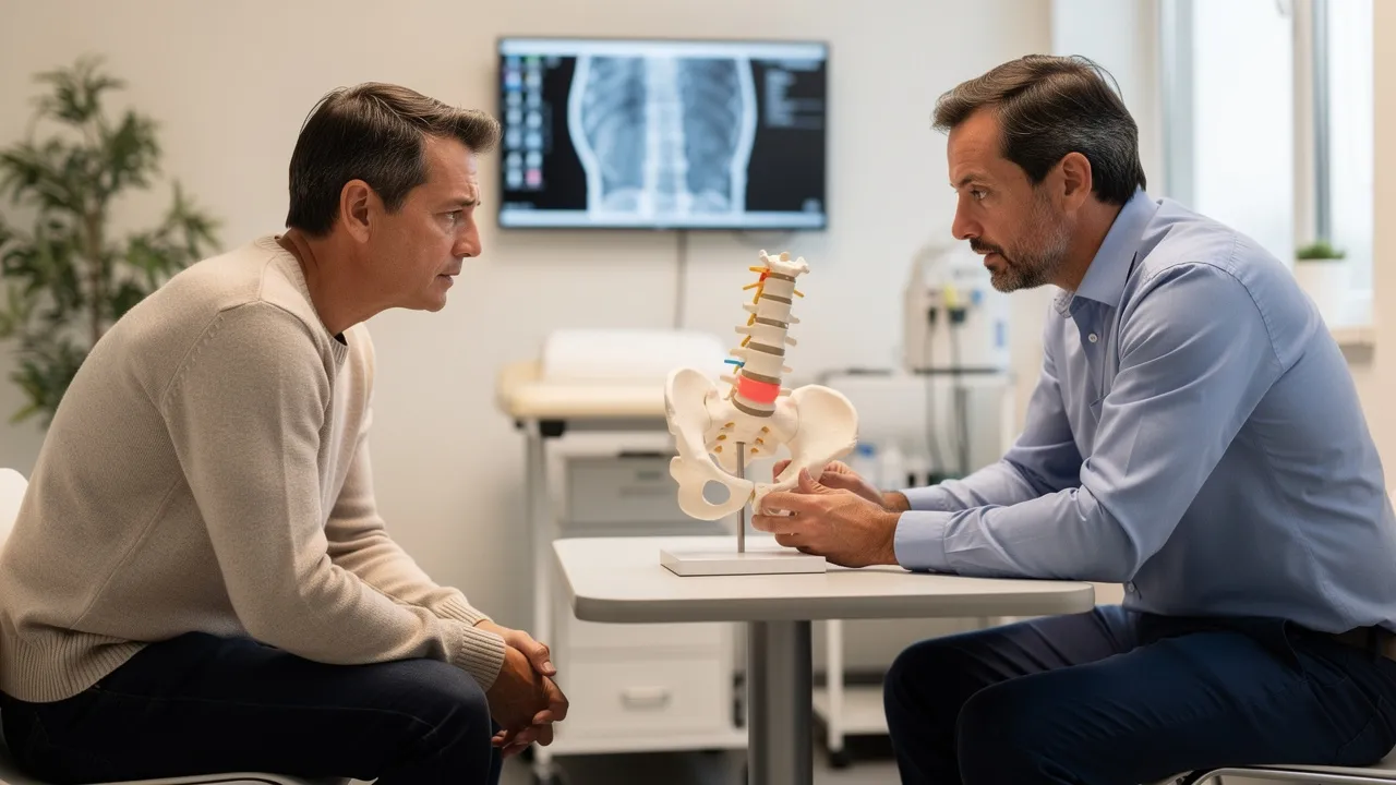
Why a slipped disc usually is not something to put back by hand
A slipped disc is usually another name for a herniated disc, not a joint that you can safely push back into place. A herniated disc occurs when part of a spinal disc bulges or ruptures and slips out of place, squeezing a spinal nerve. That pressure can produce low back pain, pain that travels into a leg, numbness, tingling, or weakness.
If you searched for how to get a slipped disc back in place, the practical answer is to focus first on symptom recovery and nerve safety rather than a hands-on repositioning technique. The affected disc may change over time, and your symptoms may improve even when the disc has not been restored to its original shape. Trying to force your spine into a position at home can also make it harder to tell whether your symptoms are changing in a concerning way.
Symptoms from a herniated disc vary. Pain may feel sharp, burning, or electric, and it may radiate into an arm or leg. Sciatica describes sharp, often shooting pain that travels from the buttock down the back of one leg when a spinal nerve is under pressure. Numbness, tingling, or weakness in the leg or foot can occur as well.
That pattern can help you describe what is happening, but it cannot identify the exact disc or establish how serious the nerve irritation is by itself. Notice where your pain travels, whether sensation is changing, and whether weakness is affecting ordinary activities. Those details are more useful for deciding what kind of assessment you need than trying to determine whether the disc has physically moved back.
Symptom improvement and disc resorption are not the same thing
Symptom improvement and disc resorption describe different changes. Symptoms can settle as nerve irritation decreases, movement becomes easier, and your back tolerates more activity. Disc resorption means that the body gradually breaks down and reabsorbs some of the herniated material. Physical therapy will not fill in a herniated disc, but the body can reabsorb herniated material over time, and physical therapy may accelerate symptom relief.
Most disc herniations improve on their own over time or with physical therapy. Improvement does not mean that every disc returns to its former appearance, and a scan does not always match how you feel. A person can have a visible disc abnormality with limited symptoms, while another person can have substantial nerve-related symptoms that need closer assessment.
Estimates for spontaneous regression vary. One estimate places regression of a herniated lumbar disc at 60% to 90% of patients, while a 2017 meta-analysis reported spontaneous resorption in two-thirds, or 66.67%, of lumbar disc herniations managed conservatively. Those figures describe changes in herniated disc material, not a promise that your pain will resolve or that your disc will return to its original position.
A separate estimate reports pain reduction within 6 to 12 weeks of conservative management in 85% to 90% of patients. That timeframe is a general reference, not a deadline for your recovery. Persistent, worsening, or function-limiting symptoms change the decision and may call for an examination rather than continued self-monitoring.
A physical-therapy source reports relief of herniated-disc symptoms for 70% to 90% of patients without surgery. That estimate belongs to the source’s reported treatment experience and should not be treated as an outcome guarantee. Physical therapy may be part of conservative care, but the right approach depends on your symptoms, examination findings, and ability to move without worsening nerve-related symptoms.

Start with measured conservative care instead of forcing the disc into position
Conservative care is a measured approach that gives symptoms room to settle while you avoid activities that make them worse. General guidance may include:
- Avoid movements that clearly increase your pain or send symptoms farther into an arm or leg.
- Avoid long periods of bed rest. Extended lying down can make joints stiff, weaken muscles, and slow recovery.
- Resume activity slowly, moving carefully when bending and lifting.
- Stay active around your home when you can do so without worsening symptoms.
- Use short walks several times a day as a general activity option when walking remains tolerable.
- Take pain medicine only within the instructions provided by a qualified clinician or the product label that applies to you.
The purpose is not to test how much pain you can tolerate. It is to keep ordinary movement gradual and observable. If an activity increases pain, numbness, or weakness rather than settling after you stop, leave that activity out and arrange an assessment if the change persists.
Activity decisions should follow your symptom response, not a fixed exercise routine. A short walk may be reasonable for one person and aggravating for another. The same applies to bending, lifting, sitting, and household tasks. Move slowly enough to notice what happens during the activity and afterward.
Do not turn general guidance into a diagnosis or a personalized exercise prescription. Specific exercises, stretches, medications, injections, or procedures require an assessment of your symptoms and relevant health factors. Stop treating home care as the complete plan if pain, numbness, or weakness is worsening or interfering more with daily tasks.
A simple daily check can keep conservative care practical:
- Record whether symptoms are staying in the back or traveling farther into an arm or leg.
- Note any new numbness or weakness and whether ordinary tasks have become harder.
- Identify which movements worsen symptoms and avoid repeating those movements.
- Reassess whether gradual activity is tolerable instead of remaining in bed for long periods.
- Arrange professional assessment when the pattern is not improving, is limiting function, or is changing in a concerning direction.
This checklist organizes what you observe. It does not determine the cause of your pain or establish that home care is appropriate.
Seek urgent care for bowel, bladder, saddle-area, or worsening nerve symptoms
Bowel, bladder, and saddle-area symptoms override routine monitoring. Seek immediate emergency medical attention for loss of bowel or bladder control, sudden urinary retention, or sudden numbness in the genital area. Sudden genital-area numbness together with sudden urinary retention and back pain is a medical emergency.
Cauda equina syndrome is rare but urgent. If it is not treated quickly, it can cause permanent nerve damage, loss of bladder or bowel control, and paralysis. It can develop rapidly, within as few as 6 to 10 hours. Do not wait to see whether these symptoms improve at home.
Worsening pain, numbness, or weakness also deserves prompt medical attention when it makes everyday activities difficult. Pay particular attention to:
- Weakness in a leg or foot that is increasing or interfering with walking or other ordinary tasks.
- Numbness or tingling that is spreading or becoming more pronounced.
- Pain that is worsening rather than gradually becoming more manageable.
- Trouble holding urine or stool, or trouble urinating when your bladder feels full.
- Loss of feeling in the inner thighs, backs of the legs, or area around the rectum, sometimes called saddle anesthesia.
These signs do not tell you the exact diagnosis, but they do establish a boundary for home monitoring. Seek medical attention instead of attempting a manual disc adjustment.
Emergency symptoms require action based on what is happening now, not on how long your back has hurt. A long history of back pain does not make new bladder changes, saddle-area numbness, or rapidly worsening weakness routine. Without prompt treatment, cauda equina syndrome can result in lasting loss of bowel or bladder function and permanent paralysis. Go to emergency care for those warning signs.

Use examination and imaging to answer the right clinical question
A spinal assessment starts with your history and a physical examination, then uses imaging when the clinical question calls for it. A clinician may ask when the pain began, where it travels, whether numbness or weakness is present, and how symptoms affect movement and daily function. The examination helps connect those details with nerve and movement findings.
An X-ray does not show a herniated disc. X-rays can answer other structural questions, but they cannot confirm the disc material pressing on a nerve. MRI can show the discs and surrounding structures more directly, although having an abnormal MRI does not by itself prove that the finding explains every symptom.
MRI becomes more relevant when findings suggest serious pathology or when neurological findings are progressively changing. MRI is the preferred diagnostic modality in many serious conditions involving cord compression or cauda equina or conus medullaris syndrome. Cited lumbar MRI criteria also identify progressive changes in objective neurological findings as a reason an MRI may be considered, with MRI without contrast rated most appropriate in that review.
That does not mean every episode of back pain requires immediate imaging. It means imaging should answer a clinical question: Is there a structural finding that matches the symptoms? Are neurological findings changing? Is there a serious condition that needs urgent attention? Discuss those questions with a qualified clinician rather than ordering or interpreting a scan on your own.
Bring focused information to an imaging or spine discussion:
- Where the pain begins and where it travels.
- Whether numbness, tingling, or weakness is present.
- Which daily activities have become difficult.
- Whether symptoms are stable, improving, or worsening.
- Any prior MRI or X-ray reports and the date of each study.
- Any bowel, bladder, or saddle-area changes that need urgent attention.
This information helps connect the scan to your current symptoms instead of treating an image as the answer by itself.
Arrange a spinal evaluation when symptoms persist or limit function
A spinal evaluation becomes a reasonable next step when symptoms persist, interfere with work or activity, or remain difficult to interpret after a period of measured conservative care. It is also appropriate when you want to discuss non-surgical options without assuming that a disc must be manually put back or that any particular treatment will be suitable.
RegenCore Method addresses degenerative disc disease, disc bulges and herniations, sciatica and radiating leg pain, facet and SI joint pain, chronic low back and neck pain, and people wanting to avoid fusion or opioids. Those are the conditions and concerns listed for our spinal disc treatment service in Palo Alto and San Francisco.
RegenCore Method evaluations include a thorough history, a hands-on orthopedic examination, diagnostic ultrasound, review of prior MRI or X-ray imaging, and a personalized treatment and recovery plan. We offer image-guided, non-surgical options for spinal disc treatment related to back and neck pain.
That process is designed to organize the next discussion around your symptoms, examination findings, and available imaging. It does not mean that a specific procedure is automatically indicated. The evaluation should clarify whether continued conservative care, further imaging discussion, or a non-surgical treatment conversation fits the situation.
Prepare for a Bay Area spinal evaluation by bringing prior imaging reports or discs when available and writing down the timeline of your symptoms. Include what you have already tried, what made symptoms worse, and whether pain or nerve symptoms have changed. If you are active in Palo Alto, Mountain View, Menlo Park, Los Altos, Sunnyvale, or San Jose, describe the activities you want to resume so the discussion can address function as well as pain.
Use urgent medical care instead of a routine appointment if you have bowel or bladder changes, saddle-area numbness, sudden urinary retention, or rapidly worsening weakness.
Understand what changes the cost of slipped-disc care
The cost of slipped-disc care depends first on which type of care you are considering. A spinal evaluation, imaging, conservative care, and surgery are different planning categories, so ask what each estimate includes before comparing amounts. A treatment conversation does not create a surgical bill, and a scan does not by itself determine whether surgery is needed.
Surgical costs can change with procedure type, geographic location, complexity, required equipment, and hospital length of stay. Insurance can change your out-of-pocket amount through the deductible, coinsurance, and whether the surgeon and facility are in network. Ask for the expected patient responsibility and the assumptions behind any estimate.
Published reference estimates provide broad surgical comparison points rather than an individual quote. A microdiscectomy is listed at approximately $15,000 to $35,000 on average, while lumbar fusion is listed at approximately $60,000 to $110,000. The difference reflects more than the procedure name: complexity, equipment, and hospital stay can all affect the total.
These figures do not establish what your care will cost in Palo Alto, San Francisco, or elsewhere in the Bay Area. They also do not establish that either procedure is appropriate for you. Surgery is considered when conservative treatments fail or when severe or worsening nerve dysfunction is present, so the clinical decision comes before treating a published price range as your plan.
Most health insurance plans cover herniated-disc surgery once it is documented as medically necessary, typically after conservative treatment has failed. Your out-of-pocket cost depends on your deductible, coinsurance, and whether the surgeon and facility are in network. Coverage rules differ by plan, and approval requirements can affect timing and responsibility.
For any proposed care, ask for a plain-language breakdown of the evaluation or procedure, imaging, facility charges, clinician charges, anesthesia when relevant, follow-up care, and expected insurance responsibility. If a treatment option is discussed without a stated price, request the amount and payment conditions before making a financial decision. RegenCore Method’s supplied service information does not establish a price, so no specific RegenCore Method fee should be assumed from the surgical ranges above.
A practical cost plan has four parts:
- Identify whether you are planning an evaluation, imaging, conservative care, or surgery.
- Separate clinician, facility, imaging, and follow-up charges where applicable.
- Check your deductible, coinsurance, network status, and any authorization requirement.
- Compare the written estimate with the care that has actually been recommended for your symptoms.
This approach keeps a surgical reference range from being mistaken for a quote and helps you ask focused financial questions before committing to care.

Choose between monitoring, evaluation, imaging discussion, and urgent care
Your next step should match the symptom pattern you have now:
- Measured monitoring: If symptoms are stable or gradually improving and no warning signs are present, avoid painful movements, avoid prolonged bed rest, and resume tolerable activity slowly. Keep tracking whether pain, numbness, or weakness is changing.
- Routine spinal evaluation: If symptoms persist, limit daily activities, or remain difficult to interpret, arrange an examination. Bring your symptom timeline and any prior imaging so the discussion can address function and nerve-related symptoms.
- Imaging discussion: If a clinician finds progressive objective neurological changes or signs that raise concern for serious pathology, discuss whether lumbar MRI is appropriate. Do not assume that an MRI is needed simply because back pain is present, and do not assume that a scan alone determines treatment.
- Urgent medical care: If you have bowel or bladder control problems, sudden urinary retention, sudden genital-area or saddle-area numbness, or rapidly worsening weakness, seek immediate emergency medical attention. Do not continue home monitoring or attempt to reposition the disc by hand.
This sequence answers the practical question behind the phrase “put the disc back”: first protect nerve function, then use symptoms, examination, imaging, and cost information to choose the next level of care.
A simple decision check can keep the order clear:
- Are emergency warning signs present? Seek immediate emergency care for bowel or bladder control changes, sudden urinary retention, saddle-area numbness, or rapidly worsening neurological symptoms.
- Are symptoms changing or limiting function? Arrange a clinical examination rather than relying on a home theory about the disc’s position.
- Is imaging being considered? Ask what question the MRI is meant to answer and how its findings relate to your examination.
- Are symptoms improving without warning signs? Continue measured activity and avoid movements that worsen symptoms while watching the trend.
- Are you comparing treatment costs? Separate evaluation and conservative-care costs from surgical reference ranges, then ask how insurance affects your responsibility.
No home maneuver can replace this sequence. A disc does not need to be manually forced into place for symptoms to improve, and symptom improvement does not remove the need to respond to new neurological warning signs.
Book a free evaluation to discuss your next step
The central decision is not how to push a slipped disc back into position. It is whether your current pattern supports measured conservative care, a spinal assessment, an imaging discussion, or urgent medical attention. Bring your symptom timeline, activity limits, prior MRI or X-ray information, and any questions about non-surgical options. A qualified clinician must decide what is appropriate for your individual symptoms and findings.
RegenCore Method offers spinal disc evaluations in Palo Alto and San Francisco that include history, a hands-on orthopedic examination, diagnostic ultrasound, review of prior imaging, and discussion of a personalized treatment and recovery plan. Use that conversation to clarify your next step without assuming a diagnosis, a procedure, or a particular outcome.
Questions to ask before choosing slipped-disc care
Will a slipped disc go back into place on its own?
Not necessarily in the sense of returning to its original shape. The body may gradually reabsorb some herniated material, and symptoms may improve as nerve irritation decreases, but those are different from manually repositioning the disc. Published estimates vary, and improvement is not guaranteed for every person.
Does a herniated disc always require surgery?
No. Many disc herniations improve with measured conservative care, such as avoiding painful movements, limiting prolonged bed rest, and gradually resuming tolerable activity. Surgery may be considered when conservative treatment fails or when severe or worsening nerve dysfunction is present. An examination is needed to determine which path fits your findings.
What should I ask about the cost of slipped-disc treatment?
First clarify whether the estimate covers an evaluation, imaging, conservative care, or surgery. Ask whether clinician, facility, imaging, anesthesia, and follow-up charges are separate, and check your deductible, coinsurance, network status, and authorization requirements. Published surgical reference ranges are not individual quotes, and the supplied information does not establish RegenCore Method pricing.
What should I bring to a spinal disc evaluation?
Bring prior MRI or X-ray reports or discs when available, a timeline of your symptoms, details about pain that travels into an arm or leg, any numbness or weakness, activity limitations, and treatments you have already tried. A Palo Alto or San Francisco evaluation may include a history, hands-on orthopedic examination, review of prior imaging, diagnostic ultrasound, and discussion of a personalized plan.
Next step
Have a question for RegenCore?
Concierge regenerative & orthobiologic orthopedics in San Francisco and Palo Alto. Research-backed, non-surgical treatment — PRP, bone marrow concentrate (BMAC), stem cell therapy, shockwave, EMTT and spinal disc care — to help the body heal itself.
Get in touch