
knee bursitis treatment
Knee Bursitis Treatment: Conservative Care, Recovery Factors, and When to Get Assessed
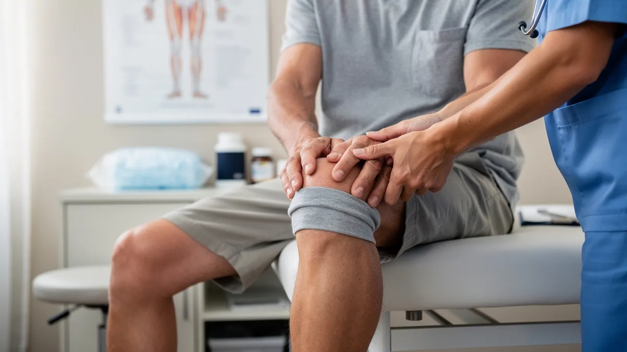
Knee swelling alone cannot confirm bursitis or rule out infection
Pain or swelling around your knee can fit superficial bursitis, but those symptoms alone do not establish the cause. Superficial bursitis is usually diagnosed from the clinical presentation, with particular attention to signs of infection. The treatment details in this guide apply most directly to superficial, especially prepatellar, bursitis; knee swelling can have other causes. Start by separating what you can observe from what still needs clinical assessment.
Signs that may occur with septic bursitis can overlap with noninfectious bursitis and, in some cases, resemble septic arthritis. Those findings are not completely reliable for identifying infection on their own, so additional diagnostic testing may be needed. Redness, warmth, tenderness, or fever can raise concern, but you cannot use their presence or absence alone to settle the diagnosis. When infection is a possibility, seek clinical assessment rather than relying on self-care to identify the cause.
For suspected uncomplicated prepatellar bursitis, conservative care commonly includes rest, ice, and activity modification. That approach does not answer every case: treatment also depends on whether infection or another underlying cause is involved. The practical distinction is whether your symptoms appear uncomplicated or leave an important question about cause. Reduce irritation when symptoms appear uncomplicated, and arrange assessment when infection is possible, the cause is uncertain, or symptoms persist or return.
Pressure, injury, inflammation, and infection can lead to bursitis
Repeated pressure is a common route to superficial bursitis. Chronic microtrauma, including kneeling on the prepatellar bursa, is the most common cause of superficial bursitis. Acute trauma or bleeding into the bursa can also be involved. If your work, exercise, or daily routine repeatedly puts pressure on the front of your knee, note that pattern. That history describes exposure, not a diagnosis. A gradual pattern associated with repeated kneeling is different information from swelling noticed after a particular injury, but neither account proves that a bursa is the source. If there is no obvious pressure pattern or event, that absence is also part of the history rather than a reason to settle on another cause. Timing, repeated contact, and a discrete injury can be described without labeling the condition. This keeps the discussion connected to the actual onset and avoids turning a common cause into an assumption about an individual knee.
Inflammatory conditions such as gout or rheumatoid arthritis are other possible causes, and infection is a separate possibility. These causes matter because changing a pressure-related activity may not address the underlying problem. Think about what happened before the swelling began: repeated kneeling, a specific injury, or a known inflammatory condition are different pieces of context to share at assessment. Do not assume that one familiar explanation accounts for every episode.
Infective bursitis has been associated with a break in the skin, trauma, and occupational risk factors; these associations have been more pronounced in prepatellar than olecranon bursitis. Staphylococcus aureus is the most common pathogen in septic bursitis, representing around 80% of cases. These details cannot tell you whether your own swelling is infected. They do make a nearby skin break or recent injury useful context to mention when you seek assessment. Septic bursitis represents around one third of olecranon and prepatellar bursitis cases. That figure describes those bursitis cases together; it is not a personal probability for someone with knee swelling, or a way to tell whether an individual bursa is infected. Assessment, rather than comparison with a group statistic, is needed to clarify the cause.
Reduce irritation with conservative measures
Conservative care for prepatellar bursitis focuses on rest, ice, and activity modification; the described approach also includes nonsteroidal anti-inflammatory medications and, in some cases, aspiration. These are options within a treatment approach, not a required checklist for every person. The cause and clinical situation affect which measures fit. In particular, aspiration is a clinical intervention, not a home-care step.
The options described also vary with the presentation. Guidance for acute traumatic or hemorrhagic bursitis includes elevation and analgesics, while other non-operative guidance for prepatellar bursitis includes compression and physical therapy. These are different sets of possible measures, not instructions to combine them. An option named for one presentation does not automatically fit another, and aspiration has a clinical role rather than being a routine self-care measure. When discussing care, it can help to ask which suspected cause and treatment goal a proposed measure addresses.
For acute traumatic or hemorrhagic bursitis, conservative treatment includes ice, elevation, rest, and analgesics. In practical terms, you can avoid adding pressure to the area and use rest and elevation while considering whether the swelling needs assessment. Aspiration may shorten symptoms in this setting, but that possibility is not a reason to attempt it yourself. A clinician determines whether an intervention is appropriate. Here, aspiration is mentioned as a possible way to shorten symptoms in acute traumatic or hemorrhagic bursitis. When infection is suspected, aspiration has a different role: obtaining fluid for examination. These are distinct reasons to consider the procedure, not interchangeable indications or a routine home-care step. A clinician can determine which question, if either, applies to the presentation.
Activities that reproduce knee pain, including kneeling, can be modified or avoided as part of non-operative care for prepatellar bursitis. Other described measures include compression over the knee, over-the-counter anti-inflammatory medications, and physical therapy to help diminish symptoms. You do not need to keep repeating an activity that clearly provokes pain to see whether your knee will tolerate it. Adjust that activity and pay attention to whether symptoms settle, stay the same, or return. Activity modification is not the same as assuming every activity must stop: the described non-operative guidance identifies painful activities such as kneeling, while recurrence is associated with activities that strain the knee. Noting whether a particular exposure appears in the history can help describe the pattern, but it cannot prove bursitis or establish a safe activity threshold. If work or exercise makes a painful exposure difficult to change, mention that constraint during assessment.
A conservative starting point for suspected prepatellar bursitis can be organized around a few choices:
- Reduce or pause painful pressure, especially repeated kneeling.
- Use rest and ice; elevation is included for acute traumatic or hemorrhagic bursitis.
- Treat medication options as part of a treatment discussion, not as an instruction that every option suits you.
- Keep aspiration and other procedures with clinical care.
- Seek assessment if infection is possible, the cause is unclear, or symptoms do not settle.
These measures come from approaches to prepatellar or acute traumatic bursitis. They are not proof that every swollen knee has bursitis, and the list does not replace assessment when another cause is a concern.

Let persistence or recurrence prompt a change in plan
Most acute cases of prepatellar bursitis respond to conservative measures. That does not mean every case follows the same course, or that improvement proves the original cause. The length of symptoms may depend in part on whether the episode is acute or chronic and whether the underlying cause is still present. Use the pattern of change—not a fixed deadline—to decide whether the current approach still seems to fit.
For mild cases, symptoms may resolve in 1–2 weeks with rest and conservative treatment. That is a possible course, not a promised recovery time or a deadline that applies to every swollen knee. The estimate concerns mild cases; it does not establish how long symptoms from infection, an ongoing cause, or a different diagnosis will last. If your symptoms are not following that kind of course, do not treat the estimate as a reason to keep waiting. Recovery descriptions differ by cause and pattern. The cited source describes chronic bursitis as persisting for several months and often recurring; it says septic bursitis may take weeks to recover, depending on infection severity and treatment effectiveness. These are descriptions of different situations, not competing deadlines or predictions for an individual. The mild-case estimate should not be transferred to persistent inflammation or suspected infection. For a longer course, reassessment can address whether the cause has been clarified and whether care is directed at it, rather than relying on elapsed days alone.
Symptoms may recur if activities that strain the knee are not modified or surrounding muscles are not strengthened. Chronic prepatellar bursitis may be managed with similar conservative therapies, and corticosteroid injections may be considered. If swelling eases and then returns, note what activity preceded the change and whether the original pressure or another cause may still be involved. Recurrence is a reason to reconsider the plan, not evidence that the same measures will be enough the next time. For chronic prepatellar bursitis, the cited guidance says conservative therapies may continue and a corticosteroid injection may be considered; another source notes that long-term physical therapy may be needed in chronic cases. These are options for clinical consideration, not evidence that every recurrence calls for an injection or that the same approach suits every cause. The guidance separately notes that recurrence is possible when surrounding muscles are not strengthened and lists physical therapy as a non-operative measure; it does not prescribe exercises or a schedule. Ask a clinician which activity plan fits your situation.
Possible infection is a reason to seek clinical assessment
Septic bursitis is more likely to involve pain or tenderness over the bursa, swelling, redness, and warmth. Fever may or may not be present, although it is more likely with infectious than noninfectious bursitis. If you notice these signs around a swollen knee, particularly with a nearby wound or skin break, arrange clinical assessment rather than treating the symptoms as routine irritation.
Redness, warmth, tenderness, swelling, and fever are clues, not a reliable home test. Clinical findings may not distinguish septic from noninfectious bursitis and can sometimes resemble septic arthritis; additional diagnostic testing may be needed. Fever is not required for infection to remain possible, and the absence of a clear sign does not establish that swelling is noninfectious. Tell the clinician about symptoms you have noticed and any recent trauma or break in the skin.
Clinical assessment may use tests to clarify the cause
Superficial bursitis is usually diagnosed from the clinical presentation, especially when the clinician is considering signs of infection. Because symptoms can overlap, the assessment may need to answer a more specific question than whether your knee is swollen: is the problem bursitis, another condition around the knee, or a potentially infectious process? Symptoms help frame that question, but they may not settle it.
Ultrasonography can help distinguish bursitis from cellulitis. That is a targeted use: it can help clarify the difference between those possibilities, rather than confirming every cause of knee pain or replacing the rest of the clinical assessment.
Blood testing and magnetic resonance imaging can help distinguish infectious from noninfectious causes of bursitis. Their role is to help clarify the question being considered, not to establish a result in advance. You can ask what uncertainty a proposed test is meant to address, so you understand how its result relates to the suspected cause.
When infection is suspected, bursal aspiration should be performed and the fluid examined. The listed tests include Gram stain, crystal analysis, glucose measurement, blood cell count, and culture. This is a clinical diagnostic step, not something to try yourself. Fluid testing can help clarify the cause when infection is a concern; the decision to perform it belongs within clinical assessment. At an assessment, it may help to understand four separate decisions: what diagnosis is being considered, whether infection remains a concern, what a proposed test is meant to distinguish, and what persistence or recurrence should prompt follow-up. These questions connect your history to the next step without asking you to interpret a test or choose a procedure independently.
Further treatment depends on the underlying cause
Treatment of prepatellar bursitis should address its underlying cause when one is identified. Examples include medical therapy for gout or antimicrobial therapy for septic bursitis. Rest, ice, and activity changes may be part of conservative care, but they do not replace treatment directed at an inflammatory cause or infection. If the cause has not been clarified, pressure reduction alone cannot answer what additional care is needed.
For chronic prepatellar bursitis, similar conservative measures may still be used, and corticosteroid injections may be considered. That wording describes a possible option, not a treatment that is appropriate for every persistent case. The underlying cause and clinical assessment shape whether an injection belongs in the discussion. Persistent symptoms call for reconsidering the cause and options, rather than treating one intervention as an automatic next step.
Incision and drainage are rarely needed for prepatellar bursitis, but may be indicated in traumatic or septic cases that do not respond to conservative measures. These are clinical interventions, not home-care choices. If your symptoms continue despite conservative care, return to the question of cause and appropriate treatment rather than attempting to drain the area yourself. These interventions are not a routine ladder of successive steps. Aspiration may be considered to examine fluid when infection is suspected or, in acute traumatic or hemorrhagic cases, for a possible effect on symptom duration. A corticosteroid injection may be considered for chronic prepatellar bursitis, while incision and drainage is rarely needed and may be indicated in certain traumatic or septic cases that do not respond to conservative care. Persistence alone does not establish which procedure, if any, is appropriate.

Use reassessment when symptoms persist or return
Persistent or recurring symptoms are a reason to reassess the working cause and care approach. Chronic prepatellar bursitis may involve continued conservative care, consideration of a corticosteroid injection, or treatment directed at a cause such as gout or infection. A change in symptoms does not identify which option fits; it signals that the original plan may need another look. Reassessment keeps the question focused on what is causing the symptoms now, not only on what was tried before.
Frequent, close follow-up as part of a structured approach to care is likely to prevent hospital admission and reduce the frequency of long-term complications. That finding describes a likelihood, not a guarantee for an individual. If you are being followed for prepatellar bursitis, keep the planned follow-up in view rather than letting unresolved or returning symptoms drift without reassessment. If the symptoms change before then, describe that change when you reconnect with the clinician.
A short symptom note can help you describe what changed. Record when swelling began, whether it followed trauma or repeated kneeling, whether the skin was broken, and whether you noticed tenderness, redness, warmth, or fever. Add whether symptoms eased and later returned, and what activity was happening around those changes. Use the notes to explain the sequence during assessment; they provide context for the clinical picture, not a way to diagnose infection at home. At follow-up, you can ask whether the working explanation still fits, whether infection or another cause remains a concern, and what change should prompt another review. The available guidance supports close follow-up but does not set one schedule for everyone, so clarify timing with the clinician rather than assuming a universal interval. This keeps the next step tied to the individual assessment, not to the notes alone.
Choose a next step that addresses the remaining uncertainty
Your next step depends on what remains unclear. If symptoms appear uncomplicated, reduce painful pressure and use conservative measures such as rest and ice. If redness, warmth, tenderness, fever, a nearby skin break, or another concern makes infection possible, seek clinical assessment because symptoms alone cannot reliably distinguish the cause. If symptoms persist or return, ask for reassessment of both the working diagnosis and the plan. A qualified professional can decide what assessment or treatment is suitable for your individual situation.
Practical questions about knee bursitis treatment
What should I do if knee bursitis makes walking uncomfortable?
The article’s guidance focuses on modifying activities that provoke pain, not stopping all movement or setting a specific walking limit. Avoid pushing through painful activity, and seek clinical assessment if symptoms persist, worsen, or raise concern about infection.
Does prepatellar bursitis always need surgery?
No. Most acute cases respond to conservative measures, and incision and drainage are rarely needed. A clinician may consider a procedure in certain traumatic or septic cases that do not respond to conservative care.
When might a clinician consider aspiration?
Aspiration is a clinical procedure, not a home-care step. It may be used to examine fluid when infection is suspected; for acute traumatic or hemorrhagic bursitis, aspiration may shorten symptoms. Whether it is appropriate depends on clinical assessment.
Next step
Have a question for RegenCore?
Concierge regenerative & orthobiologic orthopedics in San Francisco and Palo Alto. Research-backed, non-surgical treatment — PRP, bone marrow concentrate (BMAC), stem cell therapy, shockwave, EMTT and spinal disc care — to help the body heal itself.
Get in touch