
woman upper arm pain
Woman Upper Arm Pain: How to Understand the Pattern and Choose Your Next Step

Upper-Arm Pain Is a Clue, Not a Diagnosis
Upper-arm pain in a woman can begin with a shoulder-related problem even when the shoulder itself does not feel like the main source. The same general symptom can therefore point toward several different possibilities rather than one obvious answer.
The shoulder is difficult to interpret from location alone because many bony, ligamentous, muscular, and neurovascular structures contribute to its function. Pain may appear in the upper arm, while weakness or reduced use changes how the problem shows up during work, exercise, dressing, reaching, or sleep. A symptom pattern can help organize the next question, but it cannot establish your individual diagnosis.
Your first useful distinction is between a clue and a conclusion. A sore spot, a painful movement, or a feeling of weakness can tell you what to describe during an assessment; none of those features alone can identify the exact tendon, joint, muscle, or nerve involved. Start by recording where the pain sits, what movement brings it on, and what you can no longer do comfortably.
Begin With Where the Pain Is Felt
The location of upper-arm and shoulder-adjacent pain can provide a starting point, although it does not prove a cause. One orthopedic source describes rotator cuff tendonitis as the most common culprit of upper-arm shoulder pain, while also identifying overuse, accidents, and arthritis as possible contributors. Those causes can overlap, so the place where you feel pain should guide your description rather than end the investigation.
Front-of-shoulder pain may fit several shoulder-related patterns, including tendonitis involving the rotator cuff or another shoulder tendon, frozen shoulder, or bursitis. Top-of-shoulder pain may be associated with impingement, a rotator cuff injury, or an acromioclavicular-joint injury. Side-of-shoulder pain may be associated with a rotator cuff injury, frozen shoulder, or osteoarthritis. These are educational possibilities, not a way to label your condition at home.
A location map becomes more useful when you add the activity that provokes the pain. Note whether reaching overhead, lifting away from your body, reaching across your chest, or resting on that side changes the symptom. You can also note whether the pain began after a specific accident or followed repeated use. Bring those details to an evaluation instead of relying on the location alone.
For an active woman in Palo Alto or elsewhere in the Bay Area, the relevant activity may be strength training, tennis, swimming, cycling, work at a computer, or repeated lifting at home. The activity itself does not identify the injured structure. It gives you a clear way to explain when the symptom appears and how it affects your routine.
Notice What the Arm Can and Cannot Do
Arm function can add information that pain location cannot. Rotator cuff disorders include tendinopathy, partial tears, and complete tears, so the word “rotator cuff” still does not describe a single condition or a single treatment path.
Weakness deserves a precise description. Record whether you cannot raise the arm, whether you can raise it only with pain, or whether the arm feels weak after a short period of use. Also record whether your range of motion is limited by pain, stiffness, or a sense that the arm will not move farther. A clinical assessment can compare these observations with strength and range-of-motion findings.
Daily limitations can matter even when the pain number seems modest. Shoulder pain and weakness may affect daily activities, and the impact on your routine helps define what the assessment needs to clarify. Write down the tasks that have changed, such as fastening clothing, washing your hair, carrying groceries, reaching a shelf, or returning to a sport.
A painful arm does not automatically mean a tear, and preserved movement does not automatically rule one out. The broad categories overlap. Your useful next step is to describe the movement, load, and limitation clearly so a professional can test the relevant structures rather than infer a diagnosis from one symptom.
Separate Shoulder Patterns From Neck- or Elbow-Related Pain
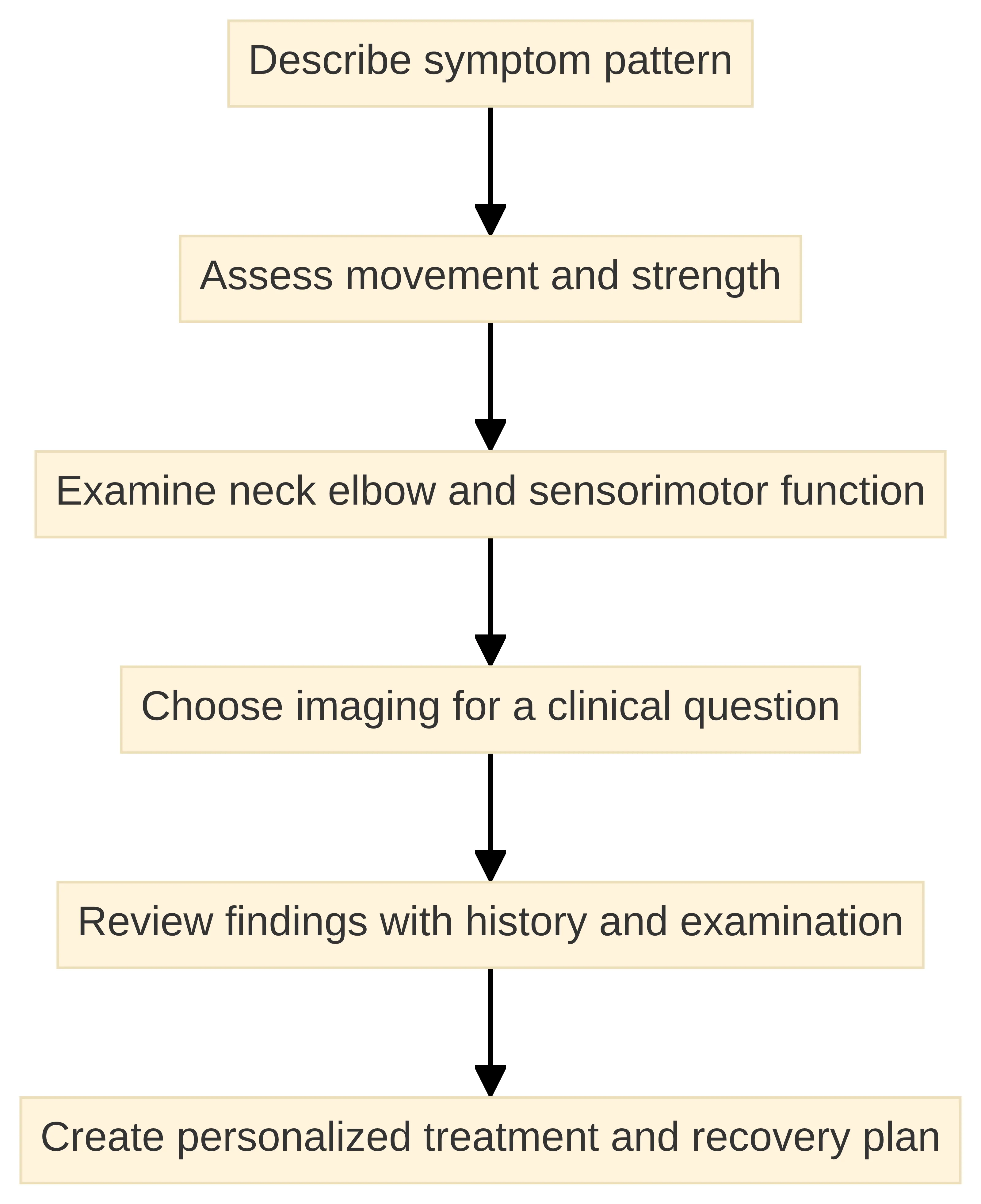
Upper-extremity pain should be assessed beyond the sore spot when the pattern does not clearly fit the shoulder. A shoulder evaluation can include inspection, palpation, range-of-motion and provocative testing, sensorimotor assessment of the arm, and evaluation of the neck and elbow. A broader examination helps separate a shoulder finding from a problem that needs attention in a neighboring region. The reason for examining neighboring regions is not to multiply possible diagnoses; it is to avoid treating the symptom’s location as proof of its source. If the neck or elbow changes the findings, that information changes the working question. If those regions do not reproduce the complaint, the shoulder remains a more focused question—but still not a confirmed diagnosis. This is why a broad first examination can be more useful than choosing a shoulder treatment from a single symptom.
The shoulder examination may also include strength testing, neurovascular assessment, and specific tests intended to reproduce symptoms and signs. These steps do not guarantee a single answer immediately, but they give the clinician a structured way to compare what hurts, what feels weak, and which movements reproduce the complaint.
A case report involving shoulder and arm pain emphasized a systematic differential-diagnosis process to support appropriate management. The practical lesson for you is simple: do not assume that an upper-arm symptom begins in the upper arm. Describe the shoulder, neck, elbow, and arm experience together, especially when the pain pattern is diffuse or difficult to reproduce.
Prepare a short map before an appointment: identify the most painful area, the movement that reproduces it, the motion that feels restricted, and any change in strength or sensation that you have noticed. That account does not diagnose the problem, but it gives the examination a specific starting point.
Let Persistence and Functional Impact Change the Next Step
Persistent shoulder-related pain deserves a different response from a brief, minor ache that has already resolved. One clinical review defines chronic shoulder pain as pain present for longer than six months and lists rotator cuff disorders, adhesive capsulitis, shoulder instability, and shoulder arthritis among common causes. The six-month definition describes chronicity; it does not mean you must wait six months before discussing pain that is limiting your life. Duration, function, and urgency answer different questions. Duration helps describe whether the problem is persistent or chronic; function shows what the symptom is costing you; urgency concerns whether waiting is appropriate. None of these dimensions identifies a tendon, joint, muscle, or nerve by itself. Keeping them separate helps you seek an assessment for the right reason instead of treating a chronic label as a diagnosis or assuming that activity limitation identifies a specific injury.
Activity-limiting pain is a practical reason to arrange professional assessment. Shoulder pain and weakness can affect daily activities, so a symptom that repeatedly changes how you work, exercise, sleep, dress, or use your arm deserves more than a location-based guess. The more specific your functional examples are, the more useful they become during an examination.
For biceps tendinopathy, one clinical review describes conservative management as potentially beginning with rest, over-the-counter pain management, and physical therapy. That approach applies to the cited condition, not to every cause of upper-arm pain. Do not assume that a self-selected treatment is appropriate until the working question is clearer.
Persistence and function can help decide when observation has stopped answering your question, but they do not determine whether a symptom is urgent. This guide cannot determine the appropriate timing of care from a page description.. This article cannot assess an individual’s urgency from a description on a page. For symptoms that are not urgent but persist or restrict your activities, arrange a professional evaluation that examines the shoulder and related regions. That separates the safety question from the later question of whether non-surgical care is worth discussing.
Know What Symptoms Alone Cannot Establish
Biceps tendinopathy is diagnosed primarily through history and physical examination, while MRI or ultrasound may help when the cause remains unclear. That principle applies to the way you interpret an upper-arm symptom: the examination and history establish the clinical question before an imaging finding is treated as the answer.
Imaging findings do not always explain symptoms or determine treatment. Anatomical variations can appear in shoulders without symptoms, and a scan may identify a tissue change without proving that the change is causing your pain. Clinicians therefore interpret imaging alongside your history and examination rather than treating a report in isolation.
In one cited study of people with atraumatic shoulder pain, routine plain radiographs altered the diagnosis in fewer than 15% of cases and changed clinical management in only 1.7% of cases. Those figures describe that study population and question; they do not mean that an X-ray is never useful. They show why the reason for ordering an image matters.
In the cited frozen-shoulder study, MRI found additional pathologies in 22% of subjects, changed treatment in 2.7% of cases, and required 37 resonance scans to identify one patient whose additional findings led to surgery. Those results reinforce a narrow point: more findings do not automatically produce a different plan. Ask what unresolved clinical question an image is meant to answer.
X-rays show injuries to the bones of the shoulder, while MRI and ultrasound provide more detailed views of soft tissues and may help identify ligament or tendon injuries. The appropriate test depends on the question being asked and the examination findings. Your symptoms can suggest what to investigate, but they cannot select a treatment or establish eligibility by themselves.
Use Pain Location to Frame the Next Step
Upper-arm pain is a symptom location, not a diagnosis. A useful first pass is to compare where the pain is felt, what movement or activity changes it, and whether the shoulder, neck, elbow, or the arm’s sensorimotor function also needs consideration. Front-, top-, and side-of-shoulder pain can suggest different possibilities, including tendon or rotator-cuff problems, frozen shoulder, bursitis, impingement, acromioclavicular-joint problems, or osteoarthritis, but location does not establish the cause. A clinical evaluation may then use inspection, palpation, range-of-motion and strength assessment, neurovascular or sensorimotor assessment, and tests that reproduce symptoms. This comparison organizes the observations for discussion; history and examination are still needed to determine what the pattern means.
Selective imaging should answer a clinical question rather than serve as a substitute for examination. X-rays can address shoulder-bone injuries, while MRI and ultrasound can provide more detailed views of soft tissues. Existing reports and images can be useful when they are interpreted alongside the symptom history and examination instead of being treated as a standalone diagnosis.
Before an evaluation, gather the date or situation when the pain began, the activities that bring it on, the movements you avoid, prior treatment such as physical therapy or injections, and any prior imaging reports. A concise symptom timeline helps connect your experience with the examination and keeps the discussion focused on function as well as pain. To make that timeline more useful, separate four kinds of information: what you felt, what you could not do, what changed it, and what has already been assessed. For example, distinguish “pain when reaching overhead” from “a rotator cuff problem,” and distinguish “could not sleep on that side” from a conclusion about the cause. Record whether an activity consistently provokes symptoms or only did so once, and whether the limitation concerns reaching, lifting, dressing, exercise, work, or sleep. Note what happened after rest, physical therapy, or an injection if any of those were tried, without treating the response as proof of a diagnosis. If you have imaging, keep the report with the date and body region; the clinician can interpret it alongside the current history and examination. This format gives the appointment a clearer starting point while preserving the difference between an observation you can report and a diagnosis that requires assessment.
Discuss Non-Surgical Options Only After the Working Question Is Clear
Non-surgical treatment options make more sense after the symptom pattern and working question are clear. Availability of an option does not establish that it fits your condition, and a treatment name does not replace examination, imaging interpretation, or a discussion of alternatives. That order creates a useful decision rule for the conversation. First identify the condition or structure under consideration; then ask which findings support it; only after that compare options, alternatives, and goals. This prevents a treatment label from becoming a substitute for a working question and makes it possible to explain why one option is being discussed while another is not. It also leaves room for the assessment to recommend a different next step when the findings do not match the intended use.
At RegenCore Method, we offer PRP for rotator cuff and shoulder tendon injuries. A discussion about PRP should therefore begin with the condition being considered and the findings that support that working question, rather than with upper-arm pain as a broad label.
At RegenCore Method, we offer BMAC for shoulder joint degeneration and severe or chronic tendon injuries. Some cartilage-injury cases may be appropriate only for select candidates. The relevant discussion is whether your examination and imaging findings fit the documented use, not whether the option sounds more advanced.
At RegenCore Method, our shockwave treatment uses a handpiece to deliver focused acoustic pulses to the injury, and most patients feel a tolerable tapping sensation. The treatment discussion still needs a defined target and a clinical reason for considering it. Upper-arm pain alone does not identify that target.
Use the evaluation to ask five practical questions: What structure or condition is being considered? Which findings support that view? What alternatives should be discussed? What role would the proposed option have in the plan? How would progress be assessed against your daily or athletic goals? Those questions keep treatment downstream of assessment without promising a particular result. Use the answers to compare the plan, not merely to collect terminology. A plan is easier to understand when it identifies the target, connects that target to examination or imaging findings, states the role of proposed care, and explains what would count as progress. If an option cannot be tied to a defined target or the findings remain unclear, ask for clarification rather than infer fit from the treatment label. This keeps the choice individualized and leaves room for the assessment to point to a different plan, rather than treating one non-surgical option as interchangeable with every other option.
Book the Next Conversation in Palo Alto
Once an urgent concern has been addressed, a non-urgent evaluation conversation can begin with a location-and-function map: note where the pain is felt, which movements reproduce it, and whether strength or sensation seems affected. Add the activity that changed—such as reaching, dressing, exercise, work, or sleep—and describe whether the change is occasional or persistent. Bring the symptom timeline and any prior treatment or imaging details so the discussion can distinguish what you have experienced from what still needs examination. These observations frame separate questions about shoulder motion, the neck, the elbow, and sensorimotor function before non-surgical options are discussed. They are preparation for an assessment, not a way to decide the cause at home.
Bring your symptom timeline, the movements that reproduce pain, the activities you have modified, and prior imaging or treatment details. A qualified professional must determine whether a particular treatment fits your condition; symptoms and scan findings alone cannot make that decision.
If Palo Alto is convenient for you, use the evaluation conversation to clarify what may be contributing to the pain, what the examination shows, and which non-surgical options are appropriate to discuss next.
Frequently Asked Questions About Upper Arm Pain Evaluation
What are common causes of upper arm pain in women?
Common causes of upper arm pain include rotator cuff tendonitis, overuse, accidents, and arthritis. These conditions can overlap, making it important to consider the specific symptoms and activities that provoke the pain.
How can shoulder-related pain present in the upper arm?
Shoulder-related pain can manifest in the upper arm due to various conditions such as tendonitis, frozen shoulder, or bursitis. The location of the pain can provide clues about the underlying issue, but it does not confirm a diagnosis.
What can I expect during an initial evaluation at RegenCore for upper arm pain?
During an initial evaluation at RegenCore, you can expect a thorough history review, a hands-on orthopedic examination, diagnostic ultrasound, and a personalized treatment plan based on your specific condition. The review of prior MRI or X-ray imaging is part of that process when those records are available, and the stated plan includes treatment and recovery planning. The purpose is to connect the symptom history, examination, ultrasound, and prior imaging rather than treat any one item as a stand-alone diagnosis.
How does the location of pain affect the evaluation process for upper arm pain?
The location of pain is crucial in guiding the evaluation process, as it helps identify potential underlying conditions. Specific movements and activities that provoke pain will also be assessed to clarify the issue.
What role does imaging play in the evaluation of upper arm pain?
Imaging, such as X-rays or MRIs, may be used to provide additional information about soft tissue injuries or bone conditions, but it is interpreted alongside the clinical examination and history.
Next step
Have a question for RegenCore?
Concierge regenerative & orthobiologic orthopedics in San Francisco and Palo Alto. Research-backed, non-surgical treatment — PRP, bone marrow concentrate (BMAC), stem cell therapy, shockwave, EMTT and spinal disc care — to help the body heal itself.
Get in touch