
shoulder pain when lifting arm
Shoulder Pain When Lifting Your Arm: How to Choose Your Next Step

Build a Movement Map Before Naming the Problem
Pain with lifting is most useful when you describe the movement precisely rather than treating it as a diagnosis. Note whether it appears during a forward reach, when the arm moves away from the body, an overhead reach, or placing the hand behind your back. Add the load, number of repetitions, and what happens as you lower the arm. Rotator-cuff-related shoulder pain can worsen when you reach up or place your hand behind your back, while pain that increases with arm use may also occur with tendonitis, bursitis, or impingement. These associations do not identify the problem by themselves.
Build the movement map before deciding what question needs answering:
- Is one direction the clear trigger, or do several lifting directions hurt?
- Does the symptom appear with an unloaded movement, a particular weight, repeated lifting, or all of these?
- Did it begin gradually, after more activity than usual, or immediately after a fall, collision, pull, or awkward lift?
- Is the shoulder merely painful, or are weakness, numbness, tingling, clicking, locking, or loss of function changing what you can do?
Use those details as a lifting ladder rather than as a home diagnostic test. First note the highest comfortable range with no added load. Then compare a familiar light task, the same task repeated, and the particular overhead or behind-the-back demand that matters to you. At each level, write down the approximate range, load, repetitions, and the point at which symptoms begin. Do not add a heavier load or more repetitions just to find a limit. If a level reproduces pain, make it a pause point and return to a less provocative task rather than repeatedly testing it. The record is useful because it compares the same demand over time; it does not show that a tissue has healed or that a condition has been ruled in or out.
Sudden, very severe shoulder pain with difficulty moving the arm or a change in shoulder shape can occur with a dislocation, fracture, or torn or ruptured tendon. That pattern belongs in a clinician’s assessment rather than in a self-diagnosis based on the lifting angle. Do not force the arm through a painful movement while recording what happened.
When those features are absent, temporarily reduce the specific painful or overhead activity and keep track of the direction and load that provoke it. A movement map can help a clinician understand the complaint, but it cannot establish a rotator-cuff problem, frozen shoulder, arthritis, or another named condition. Its purpose is to make the next clinical question more precise.
Start With the Exact Lifting Problem
Pain with lifting becomes more useful when you describe the movement precisely. “My shoulder hurts when I raise my arm” can mean several different patterns. A clinician can work with a more specific account.
Start by recording these details:
- Direction: Does pain begin when you reach forward, move your arm out to the side, reach overhead, or place your hand behind your back?
- Position: Does the pain stay at the shoulder, travel into the upper arm, or extend toward the neck or shoulder blade? Rotator-cuff-related pain can spread into the upper arm and may extend toward the neck or shoulder blade.
- Timing within the movement: Does the pain begin immediately, appear only as the arm gets higher, or continue after you lower it?
- Load: Does an empty-handed movement hurt, or does the problem appear when you carry, push, pull, throw, or perform another demanding task?
- Onset: Did the symptom start suddenly after a fall, collision, lift, or other event, or did it build gradually?
- Function: What can you no longer do comfortably? Examples include reaching a shelf, putting on a shirt, fastening clothing, carrying equipment, swimming, serving, lifting at work, or returning to a training movement.
- Companions: Have you noticed stiffness, tingling, numbness, weakness, clicking, locking, or a visible change in shape?
Pain that becomes worse as the arm moves away from the body, including during reaching up or placing the hand behind your back, can fit a rotator-cuff-related pattern. Pain that is worse while using the arm can also occur with tendonitis, bursitis, or impingement. These descriptions help frame the examination; they do not establish which condition you have. “I cannot serve” gives a different practical question from “I cannot reach a high shelf,” even if both movements reproduce pain. A useful description connects the symptom to the demand that matters to you.
Compare Broad Cause Categories Behind Painful Elevation
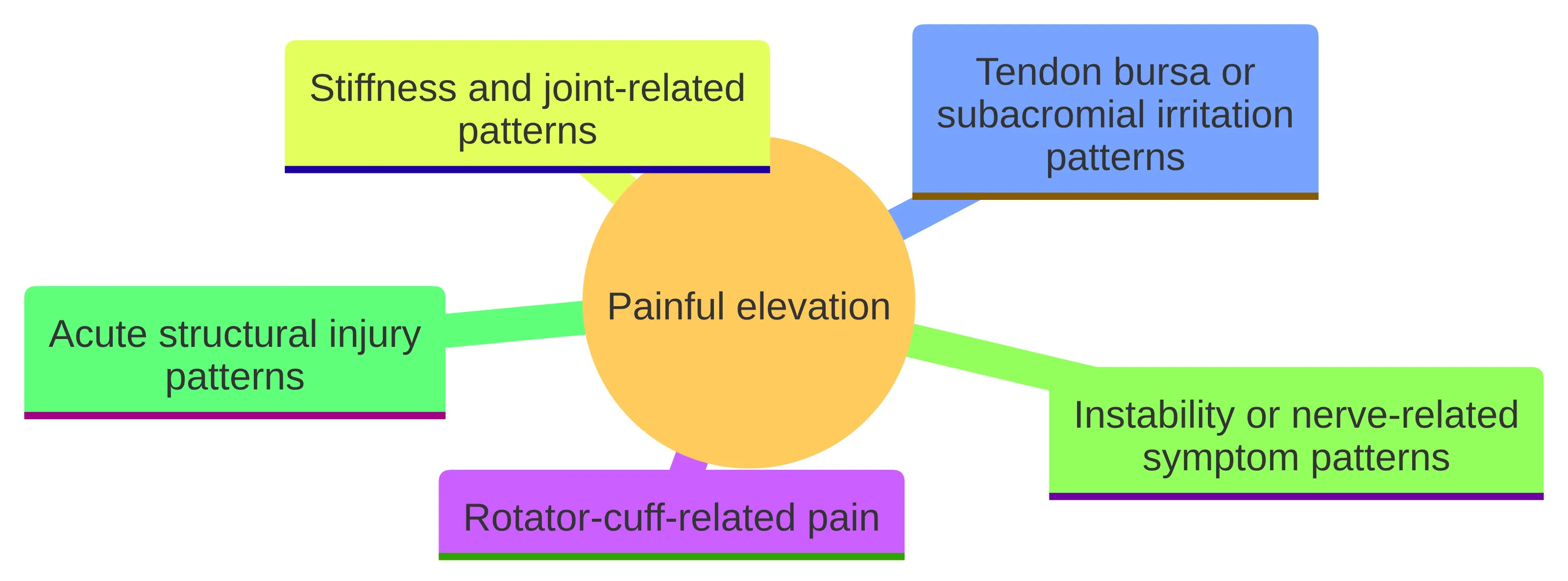
Painful elevation can fit several broad categories. Use them to organize your account, not to assign yourself a diagnosis.
Rotator-cuff-related pain can affect the shoulder and upper arm, sometimes extending toward the neck or shoulder blade. Reaching up or placing your hand behind your back may worsen it. This category describes a symptom pattern and does not tell you whether a tendon is irritated, strained, or torn. NHS symptom guidance lists tendonitis, bursitis, and impingement among possible associations with that pattern. The same movement can therefore occur across more than one category. Frozen shoulder and arthritis are among the possible associations listed for persistent pain and stiffness. Pain during lifting alone does not establish either one. Those symptoms can occur with shoulder instability, sometimes in connection with hypermobility, but they do not establish that explanation. A clinician can then determine which tissues, joints, or other contributors need assessment. That assessment is not limited to matching a symptom to a familiar label. It can consider whether the complaint is primarily painful, stiff, weak, unstable, or difficult to move; whether the onset followed an event; and whether the shoulder’s function is changing. The categories overlap, so the same person may describe more than one feature. For example, pain during use and reduced motion may coexist without showing whether the main issue involves a tendon, bursa, joint, or another structure. Treating the categories as prompts for better questions preserves that uncertainty while still making the next step more concrete.
Compare Direction, Load, and Onset
Make the lifting record comparable rather than exhaustive. Start with an unloaded version of the movement, staying within a range that does not require forcing the arm. If that movement is relevant to daily life, note the point where symptoms start. On another occasion, compare a familiar light task, then the same task for repeated movements, and finally the work, sport, or overhead demand that matters to you. Keep the direction, approximate load, repetitions, and symptom onset visible for each level. This is a way to describe changing capacity, not a strength test or a home diagnosis.
A practical rule keeps the comparison from becoming a challenge: do not increase load, range, or repetition to chase a limit. When one level reproduces the symptom, record that level and stop escalating that session. A level that remains comfortable is simply the next observation; it is not evidence that the shoulder is healed. If pain appears earlier, the tolerated load falls, or the task remains restricted, preserve that change in the record. If the same ordinary task remains painful, repeatedly brings the symptom back, or continues to limit work, household activity, exercise, or sport, the comparison tells you what to bring to an evaluation to answer the clinical question.
The first episode still matters. Note whether symptoms built after more activity than usual or began after a fall, collision, forceful pull, or awkward lift. Rotator-cuff-related pain may begin slowly or relatively quickly after a more demanding activity, but timing describes the account rather than identifying the tissue involved.
Compare the original map with what you can do now:
- Can you complete basic tasks, or does the same direction repeatedly stop you?
- Has the load, reach, or number of repetitions you tolerate become smaller?
- Are you compensating with your neck, back, or opposite arm?
- Have you changed work duties, exercise, sport, or sleep positions because of the shoulder?
- Is weakness, numbness, tingling, clicking, or locking changing the map?
A gradual onset is not automatically a reason to ignore the symptom, and an injury history is not a diagnosis. If the shoulder is changing what you can do in Palo Alto, Mountain View, Menlo Park, Los Altos, Sunnyvale, San Jose, or elsewhere in the Bay Area, describe the affected task and load directly. Function turns a list of symptoms into a question a clinician can assess.
Recognize What Symptoms Cannot Establish Alone
A painful lifting pattern cannot tell you by itself whether the problem is in a tendon, bursa, joint, bone, nerve, or more than one structure. Several conditions can produce pain with arm use, and a single associated symptom can have more than one possible explanation.
For example, pain when reaching overhead can occur with rotator-cuff-related shoulder pain. Pain that worsens while using the arm can also occur with tendonitis, bursitis, or impingement. Persistent pain and stiffness can occur with frozen shoulder or arthritis. Tingling, numbness, weakness, clicking, or locking can occur with shoulder instability, although those symptoms do not establish a diagnosis. A label can influence which activities you avoid, which exercises you try, and whether you delay an evaluation. Let the symptom pattern guide the questions you bring to a qualified clinician instead.
The same boundary applies to imaging. An image can answer a specific structural question, but deciding which test is appropriate depends on the clinical context. You do not need to decide that an MRI, ultrasound, or injection is the answer before the shoulder has been assessed.
Modify the Specific Lift That Provokes Pain
For a pattern without the sudden, very severe pain, marked difficulty moving the arm, or change in shoulder shape described earlier, apply the lifting ladder to the provoking task rather than testing the whole shoulder:
- Reduce the specific reach or lift instead of repeatedly checking the painful limit.
- Keep the load and repetition below the point at which symptoms begin rather than escalating to find the threshold again.
- Treat a comfortable movement as a separate comparison; do not assume it clears a painful overhead or behind-the-back demand.
- Do not force a range of motion or use a painful exercise as a self-test.
- Before starting shoulder exercises, discuss them with a doctor or physical therapist. AAOS advises that conversation before attempting them.
If the same ordinary task remains restricted, the symptom returns whenever normal activity resumes, or function continues to fall, keep the record at that level and arrange an evaluation rather than progressing the ladder. The purpose is to describe the boundary of the problem without turning activity modification into a diagnosis or a personal treatment plan.
That information supports activity modification and supervised exercise as topics for discussion; it does not turn a lifting pattern into a diagnosis or a personal treatment plan.
Record what changes when the direction, load, or repetition changes. A short-term reduction in pain does not establish that the shoulder has healed, and continued pain does not by itself establish a need for an injection or surgery.
Use Function to Decide When the Map Needs a Clinician
Read the record by comparing the same ladder level with the same level later, rather than by treating one pain score as the whole result:
- Does the same load and range remain available, become more limited, or bring symptoms on sooner?
- Can you complete the same number of repetitions, or does the symptom interrupt the task earlier?
- Has a work, household, exercise, or sport demand become available, stayed restricted, or been lost?
- Are weakness, numbness, tingling, clicking, or locking adding a new feature?
- Is a previously tolerable direction now provoking symptoms?
A change in recorded capacity is information about the task, not proof of healing or a diagnosis. If the record shows persistent restriction, repeated return of the symptom with ordinary activity, or worsening function, it supports arranging a clinical assessment rather than raising the load or range to obtain a clearer result.
Persistent pain and stiffness over months or years can occur with frozen shoulder or arthritis, while pain that remains worse during arm use can occur with tendonitis, bursitis, or impingement. Those associations do not set a personal timetable, but they explain why a symptom that does not change with activity modification deserves a clinical question rather than repeated experimentation.
A clinician’s evaluation becomes the appropriate next question when the problem persists, returns whenever normal activity resumes, or continues to limit function. Include the direction, load, onset, and associated symptoms in that account. Keep the record simple enough to use: note the task, direction, approximate load, repetitions, and what you could do afterward. This is not a diagnostic test or proof of improvement. It provides a consistent comparison for the clinician and may show whether the problem is stable, changing, or linked to one demand. If function is worsening or a new associated feature appears, do not treat an earlier period of improvement as evidence that the current pattern is unchanged. Bring the updated map to the evaluation.
Bring the Movement Map Into the Evaluation
Bring the lifting-ladder record to the evaluation instead of presenting only a pain location. Show the comparison that matters most:
- the direction of the lift: forward, away from the body, overhead, or behind the back;
- the unloaded and loaded versions of the task, including approximate load, repetition, and point at which the symptom begins;
- whether the threshold changed after activity was reduced or normal activity resumed;
- whether onset was gradual or followed an injury;
- where the pain travels and whether function has changed; and
- whether stiffness, weakness, numbness, tingling, clicking, locking, or a visible change accompanies it.
This gives the clinician a concrete account of what the shoulder can and cannot do, while leaving the cause open for history and examination.
The evaluation can then relate that account to the activity that matters to you. RegenCore Method’s evaluations include a treatment and recovery plan personalized to the findings. The purpose is to relate findings to an individual plan rather than treat every painful lifting pattern as the same problem.
Imaging answers a narrower question after the clinical account has been established. X-rays show shoulder-bone injuries, while MRI and ultrasound provide more detailed soft-tissue images than X-rays; MRI may help identify ligament and tendon injuries. The imaging choice depends on what the assessment is trying to establish, not simply on which lifting angle hurts.
Describe the activity you want to resume, whether that is tennis, climbing, swimming, strength training, or overhead work. A direction-and-load map gives the clinician a practical starting point for discussing conservative care, rehabilitation, further assessment, or another option.
A practical evaluation sequence keeps those discussions connected rather than treating each test or treatment as a separate answer:
- Clarify the clinical question. Is the main uncertainty about the painful movement, a change in strength or function, stiffness, the effect of an injury, or another associated symptom?
- Relate the history to the examination. The onset, load, direction, and functional change provide context for what the hands-on orthopedic examination needs to assess.
- Use imaging for the question that remains. RegenCore Method states that its evaluations include diagnostic ultrasound and review of prior MRI or X-ray imaging. Those imaging inputs can contribute to the assessment, but neither a scan nor an ultrasound finding establishes the cause of pain by itself.
- Connect findings to a plan. The personalized treatment and recovery plan should identify the activity goal, the care options being discussed, and what uncertainty or finding would lead to reassessment.
If you have prior imaging, bring the report and images when available, and explain when they were obtained and what has changed since then. Ask whether the current movement pattern or functional limitation changes how those records should be understood. This approach helps distinguish an imaging result that informs the next decision from one that simply adds a label without clarifying what to do.
Use Imaging to Answer the Clinical Question
Imaging should answer a clinical question rather than substitute for one. Different tests show different aspects of the shoulder, and the appropriate sequence depends on the symptom history, examination, and concern being investigated.
For chronic shoulder pain, the American College of Radiology narrative says that most literature supports radiographs as the initial imaging study. The same narrative reports insufficient evidence to support MRI with or without intravenous contrast or MR arthrography as the initial imaging study for chronic shoulder pain. That guidance applies to the initial evaluation of chronic shoulder pain; it does not mean that MRI is never appropriate. – Ultrasound and MRI provide more detailed soft-tissue images than X-rays.
- MRI may help identify injuries involving ligaments and tendons.
- Other tests may be considered when the clinical question involves a different structure or function.
These distinctions help explain why a clinician may not order the most advanced-sounding test first. X-rays, MRI, and ultrasound are not interchangeable, and a finding on an image still needs to be interpreted alongside your symptoms and examination. RegenCore Method states that its evaluations include review of prior MRI or X-ray imaging as part of the assessment. That review can keep the discussion connected to what has already been investigated instead of treating the image as an isolated answer. If the answer is unclear, ask which finding would change the next step. That keeps imaging connected to care rather than turning it into a search for an explanation without an action. One way to keep the sequence clear is to ask what structure or question is under review before discussing the test. If the concern is an injury to the bones, X-rays can show shoulder-bone injuries. If the concern involves soft tissue, MRI and ultrasound provide more detailed images than X-rays, and MRI may help identify ligament or tendon injuries. These are capabilities of the tests, not a reason to select one without an examination. For chronic shoulder pain, the ACR statement about radiographs as the initial study likewise describes an initial imaging approach, not a promise that the first image will settle the cause. Ask what the clinician is looking for, what the result could change, and how the result will be interpreted with the history and examination. This makes an imaging conversation more useful than choosing a test because it sounds more comprehensive. It also helps prevent a prior scan from becoming the entire explanation when the current movement, function, or onset has changed.
Match Non-Surgical Discussions to the Finding
Non-surgical care is not a single treatment and it is not automatically the right answer for every painful lifting pattern. The sensible sequence is to establish what is being treated, what your shoulder needs to do, and which options fit the findings before discussing a procedure.
- Is the immediate plan activity modification, rehabilitation, imaging, an injection discussion, or another step?
- What would make the plan change?
- How will the plan relate to the work, sport, or daily activity you want to resume?
- What uncertainty remains about suitability, expected course, or alternatives?
RegenCore Method can guide patients across the full spectrum of care and reserve surgery for when it is considered the right answer. That describes RegenCore Method’s evaluation and care approach, not a prediction about what you will need.
Keep a non-surgical discussion tied to the finding being addressed. An available option is not automatically appropriate for a painful lifting pattern, and a conversation about PRP or BMAC is not an individualized recommendation. That distinction also applies to the difference between a service being offered and a treatment having condition-specific support. The lifting record gives that conversation a concrete starting point. Ask the clinician to look at the range, load, repetitions, and symptom threshold together, then explain which finding, if any, makes a non-surgical option worth discussing. A procedure should not be selected simply because it is available or because the painful movement has a familiar label. Ask what the option is intended to address, what conservative path remains, and what observation would lead to reassessment.
For example, a person whose record shows pain only after a recent increase in overhead demand can bring the exact task, load, and point of onset to a discussion about activity modification or supervised rehabilitation. A person whose record shows a sudden event, a persistent functional loss, or a different associated symptom may need the assessment to answer another question first. These examples organize the conversation; they do not identify a condition or select a treatment. The useful distinction is not “non-surgical” versus “surgical” in the abstract. It is whether the proposed step connects to the individual findings and the activity goal, with alternatives and remaining uncertainty stated plainly. That keeps availability separate from suitability and avoids turning a treatment discussion into a promise about pain, tissue repair, or recovery.
Separate Availability From Evidence for PRP and BMAC
PRP and BMAC belong in a clinical discussion after the shoulder problem has been assessed. Their availability does not establish that either option is suitable for your symptoms, and the evidence is not interchangeable between the two.
RegenCore Method states that it offers PRP for conditions that commonly include rotator-cuff and shoulder tendon injuries. RegenCore Method also states that it offers BMAC for conditions that commonly include shoulder joint degeneration and severe or chronic tendon injuries. Those are descriptions of the conditions the practice commonly addresses; they are not a determination that either treatment fits your shoulder. It also describes the PRP findings as heterogeneous, says effects are generally modest, and notes that some trials show no advantage over saline or corticosteroid. A larger evidence base does not mean a predictable result for an individual shoulder. It reports that standalone shoulder BMAC randomized trials are essentially absent. It also reports that no published randomized trial directly compares PRP with BMAC for a shoulder disorder. That means you should be cautious about claims that one option is established as the better choice for your lifting pain. Those statements place both options downstream of assessment and informed discussion rather than in the position of automatic first-line care. – Is the proposed use an injection on its own, an adjunct to another procedure, or part of a broader plan?
- What shoulder-specific evidence supports that use?
- What uncertainty remains about benefit, alternatives, cost, and recovery demands?
- What would the plan be if you chose rehabilitation or continued conservative care instead?
- How would progress be assessed against the activity you want to resume?
A treatment conversation is more useful when it explains why an option is being considered and what would count as meaningful progress. It should not turn a symptom pattern into a promise.
Turn the Symptom Account Into the Next Clinical Question
Before an appointment, turn your experience into a short brief. The brief does not need a self-diagnosis. It needs enough detail to help the clinician decide what to examine and what question to answer first.
Write down:
- The trigger: “Pain begins when I reach forward,” “pain appears as I lift out to the side,” or “pain starts when I put my hand behind my back.”
- The location: shoulder, upper arm, neck, or shoulder blade, including whether the pain spreads.
- The onset: gradual, sudden, or linked to a specific fall, lift, collision, or unusually demanding activity.
- The function change: the work task, household task, exercise, or sport movement you cannot perform normally.
- The associated symptoms: stiffness, weakness, tingling, numbness, clicking, locking, or a change in shoulder shape.
- The interim response: which activities you reduced and whether the pattern improved, remained unchanged, or returned when you resumed them.
- The records: prior X-ray or MRI reports and images, if available.
Then turn the brief into a direct question: “What needs to be established before I decide whether to continue activity modification, begin clinician-guided rehabilitation, pursue imaging, or discuss a non-surgical procedure?”
This approach keeps the conversation tied to your actual goal. If your goal is returning to tennis, lifting, swimming, climbing, or overhead work in the Bay Area, include the movement, load, and frequency that matter. Rotator-cuff-related pain can worsen with reaching up or placing the hand behind the back, and instability-associated symptoms can include tingling, numbness, weakness, clicking, or locking, so those details may change the evaluation question. They still do not establish a diagnosis by themselves. Before the appointment, decide which uncertainty matters most: identifying what movement is involved, understanding why function changed, choosing an interim activity boundary, or comparing a non-surgical option with rehabilitation. Lead with that uncertainty rather than with a treatment you have encountered elsewhere. If you are considering PRP or BMAC, ask the clinician to connect the proposal to the specific finding and explain what evidence applies to that shoulder problem. If the main concern is returning to sport or work, ask how progress would be judged in that activity rather than by pain alone. These questions do not predict the outcome; they make the discussion more transparent and leave room for the plan to change after examination or imaging.
Use Your Movement Map to Choose the Next Question
Pain during arm elevation is a useful starting clue, not a diagnosis. First separate sudden severe pain, marked difficulty moving the arm, or a change in shoulder shape from a pattern that can be observed while you reduce painful activity. Then describe the exact movement, onset, associated symptoms, and effect on your work or sport. If the problem persists, recurs, or continues to limit function, an evaluation can turn uncertainty into a specific clinical question.
We discuss non-surgical options only in relation to the individual findings and the activity you want to resume. PRP and BMAC remain discussion points rather than automatic answers: current review evidence describes more shoulder-specific research for PRP but heterogeneous and generally modest effects, while shoulder BMAC evidence is sparse.
A qualified clinician must determine individual suitability, interpret warning features, and decide whether a treatment or imaging study fits your situation. Your notes can turn the visit into a focused comparison: what the history suggests, what the examination needs to clarify, whether imaging would change the plan, and which conservative or non-surgical options remain open. That sequence keeps the decision tied to evidence about your shoulder rather than to the name of a treatment. It also leaves room for the plan to change if function, associated symptoms, or imaging findings differ from the initial account.
What Should You Ask Before Discussing Non-Surgical Shoulder Care?
How can I describe my shoulder pain to a clinician?
When describing your shoulder pain, include details such as the direction of movement that causes pain, the location of the pain, when it started, any changes in function, and any associated symptoms like weakness or numbness.
When should I consider seeking a professional evaluation for my shoulder pain?
Consider seeking evaluation if your shoulder pain persists, recurs with normal activity, or limits your function.
What initial steps can I take to manage shoulder pain when lifting my arm?
You can manage shoulder pain by temporarily reducing or avoiding activities that provoke pain, keeping track of your symptoms, and consulting a clinician before starting any exercises.
What non-surgical options are available for shoulder pain?
Non-surgical options may include physical therapy, activity modification, and possibly injections like PRP or BMAC, but these should be discussed after a thorough evaluation.
Next step
Have a question for RegenCore?
Concierge regenerative & orthobiologic orthopedics in San Francisco and Palo Alto. Research-backed, non-surgical treatment — PRP, bone marrow concentrate (BMAC), stem cell therapy, shockwave, EMTT and spinal disc care — to help the body heal itself.
Get in touch