
what can cause knee pain without injury
What Can Cause Knee Pain Without Injury? A Symptom-Pattern Guide
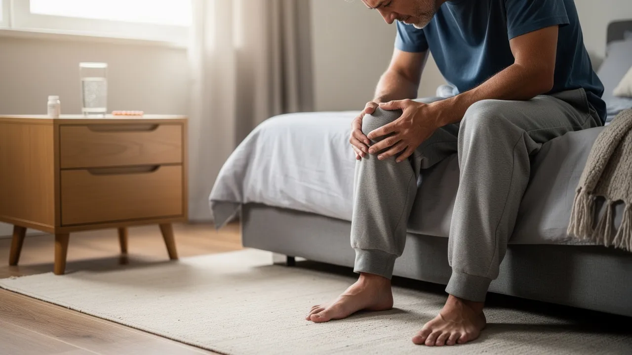
Begin with the pattern, not a single suspected cause
Knee pain can have several possible explanations. Knee pain is common and can have many possible causes, so the absence of a clear twist, fall, or impact does not identify the source by itself.
The useful starting point is the pattern around the pain. Notice whether symptoms developed gradually or suddenly, where the pain sits, what activities bring it on, whether the knee is swollen or warm, and whether the joint still moves and bears weight normally. Those details can help sort broad possibilities and determine how quickly you need an evaluation.
A knee pain evaluation considers age, pain location and timing, pain quality, mechanical or systemic symptoms, swelling, trauma, and relevant medical or surgical history. You can use the same categories to organize what you are noticing in Palo Alto, Mountain View, Menlo Park, Los Altos, Sunnyvale, San Jose, or elsewhere in the Bay Area.
Knee pain affects approximately 25% of adults, and knee pain accounts for nearly 4 million primary care visits annually. The 25% figure measures adults, while the visit figure measures annual primary-care visits rather than the number of people with a particular diagnosis. These figures describe how often knee pain appears in clinical care, not what is causing your symptoms.
The timing, location, triggers, swelling, and function of knee pain help separate a routine evaluation from a situation that needs faster attention. The timing, location, triggers, swelling, and function of knee pain help organize what to report to a qualified professional. Locking, giving way, or inability to bear weight changes the practical next step even when you cannot name the cause.
Use these observations to describe your symptoms clearly. Do not use them to diagnose yourself.
Gradual pain and stiffness often point toward joint or cartilage problems
Knee osteoarthritis develops as cartilage in the joint breaks down and may cause stiffness, pain, reduced movement, or grating and crackling sensations. Osteoarthritis pain usually progresses slowly and is more persistent than acute pain from repetitive activity. You may notice discomfort during ordinary weight-bearing, a gradual change in how far the knee moves, or a grinding sensation when the joint bends and straightens.
Knee osteoarthritis can cause pain, swelling, localized tenderness, loud clicking, joint deformities, and stiffness lasting no more than 30 minutes after waking. The 30-minute description refers to morning stiffness in people with knee osteoarthritis; it is a symptom pattern, not a threshold that confirms the condition.
Knee osteoarthritis is more common among people over 50, and women are more susceptible than men. The age figure describes a population pattern, not a rule about your knee. A personal history of knee injury or surgery and a family history of knee osteoarthritis or joint replacement are established risk factors, while other associated factors include overweight or obesity, repetitive occupational knee stress, and certain alignment patterns.
These associations can help you give a clinician useful background, but they do not establish that osteoarthritis is present. Someone younger than 50 can have knee pain from several other causes, and someone over 50 can have a different problem with a similar ache or stiffness.
A gradual pattern matters more than any single sound or sensation. Persistent discomfort, reduced movement, swelling, clicking, or stiffness after waking can occur with knee osteoarthritis, but these features can overlap with other knee conditions. Record how long morning stiffness lasts, whether both knees are affected, and whether ordinary activities are becoming harder.
Risk factors are context, not proof. A prior injury, family history, age, body size, work demands, or alignment pattern can make osteoarthritis part of the clinical conversation without identifying it as the cause of your pain.

Pain around the kneecap can follow repeated loading
Patellofemoral pain can cause gradual, diffuse aching at or behind the kneecap, often worsened by stairs, squatting, running, jumping, or prolonged sitting. Repetitive running, jumping, squatting, kneeling, or similar activities can stress the patellofemoral joint and contribute to anterior knee pain. That pattern can appear in active adults who run on Bay Area trails, cycle, train in a gym, play recreational sports, or spend long periods seated at work.
Patellofemoral pain is common in young athletes and may remain common through adulthood. A reported incidence as high as 6% applies to patellofemoral syndrome or idiopathic anterior knee pain; that percentage does not measure all knee pain or show that your pain has that cause.
Pain around the kneecap, stiffness after sitting, pain with the knee bent, and discomfort that eases after walking for a minute or two can occur with patellofemoral pain or early arthritis. Popping, clicking, or grinding when you rise, along with a knee that feels tight or initially will not fully straighten, can accompany anterior knee pain patterns.
Pain reproduced during a squat is the best available clinical test described for patellofemoral pain, but no definitive test exists and other causes should be excluded. Treating a squat response as a diagnosis can send you down the wrong path, especially if swelling, locking, instability, or systemic symptoms are also present.
Activity-related knee pain can build without one memorable incident. Repeated stairs, running, jumping, kneeling, or squatting may load the joint behind the kneecap, while active adults may also experience overuse problems such as pes anserine bursitis or medial plica syndrome.
The practical clue is the relationship between activity and symptoms: record which movement brings on pain, where you feel it, and whether it settles afterward. A consistent trigger can guide a qualified evaluation, but it cannot identify the involved structure by itself.

Localized tenderness may involve a tendon or bursa
Knee tendinitis affects the tendons that connect muscle to bone; overuse or injury can make these tissues inflamed and painful. Tendinitis often causes a dull ache that increases with use, and swelling may also occur. A pain pattern that follows repeated loading and feels more localized than a deep, diffuse joint ache can fit a tendon-related possibility, but symptom location alone cannot confirm it.
Patellar and quadriceps tendinosis are among the possible causes of chronic anterior knee pain associated with overuse. The word “possible” matters: a tendon, the joint behind the kneecap, a bursa, or another structure can produce pain in a nearby area.
Knee bursitis is inflammation of a bursa that may follow repetitive pressure or trauma and can cause localized swelling and tenderness at the front of the knee. Repeated kneeling, pressure, or activity can make the area feel sore to touch.
Other possible causes of chronic anterior knee pain associated with overuse include fat pad impingement and plica syndromes. Active patients are more likely to have overuse injuries such as pes anserine bursitis and medial plica syndrome. These broad categories can help you describe the pattern; they do not tell you which tissue is involved.
Localized tenderness is useful information when you describe it precisely. Note whether the sore area is at the front of the knee, over a tendon, or near a superficial bursa, and whether use increases the ache or direct pressure increases tenderness.
A localized pattern does not automatically mean the problem is minor. New swelling, increasing pain, reduced movement, or trouble bearing weight should move you from observation toward qualified evaluation.
Sudden hot swelling changes the category of concern
Gout can cause rapid-onset severe pain, swelling, warmth, and redness in the affected joint, including the knee. Gout pain may peak within 4 to 12 hours after onset and may be worst within 8 to 12 hours. Discomfort may last from a few days to a few weeks, and moving the affected joint may become difficult.
Rheumatoid arthritis can cause pain, swelling, stiffness, inflammation, and loss of function, often affecting corresponding joints on both sides of the body. A pattern involving both knees or other matching joints can be relevant background, but it does not distinguish rheumatoid arthritis from every other inflammatory condition.
Acute infectious arthritis can develop over a few hours to a few days and cause moderate to severe joint pain, warmth, tenderness, swelling or effusion, and restricted active and passive movement, usually in one joint. Septic arthritis is a joint infection that can evolve over hours or days and rapidly destroy joint structures; diagnosis requires synovial fluid analysis and culture.
Many, but not all, people with acute infectious arthritis are febrile, and systemic symptoms may occasionally be minimal or absent. A knee that becomes hot, red, swollen, and painful therefore deserves urgent attention even if you do not feel generally ill. Do not wait for a remembered injury before taking that pattern seriously.
Sudden warmth and swelling can occur with gout, inflammatory arthritis, or infection, so the appearance alone cannot tell you which condition is present. Rapid severe pain, restricted movement, redness, or swelling should be treated as a change in urgency rather than as a reason to choose a home remedy.
Seek urgent medical attention when a hot or red swollen knee occurs with fever. Seek urgent attention for a rapidly painful, warm, swollen knee even without fever when movement becomes restricted or the overall pattern is changing quickly.

Mechanical symptoms within the knee can change the next step
A meniscal tear may cause catching or clicking, locking or giving way, joint-line tenderness, loss of extension when the knee is locked, and effusion in 24 to 48 hours. Popping or grinding alone is less specific, but locking, giving way, or an inability to fully straighten changes the need for evaluation.
Over 100 different types of arthritis can affect the knees. That broad figure helps explain why “arthritis” is not a single symptom pattern and why mechanical symptoms should not automatically be labeled as ordinary wear. A knee that catches, locks, or repeatedly gives way needs assessment rather than repeated home testing.
Function can be more useful than the sound a knee makes. Note whether you can fully straighten the joint, walk normally, bear weight, and move through ordinary activities without giving way. A click without functional change has a different practical meaning from a knee that locks or repeatedly becomes unstable.
Describe the change plainly. “My knee catches before it straightens” or “my knee gives way while walking” gives a qualified professional information that a label such as “meniscus problem” does not.

Use patterns as clues, not a home diagnosis
Knee symptoms can overlap among osteoarthritis, overuse problems, inflammatory conditions, crystal arthritis, infection, and mechanical disorders. Because symptoms can overlap among knee conditions, a symptom pattern alone cannot establish your diagnosis.
History and examination findings can help distinguish possible causes. Imaging is not a substitute for describing the onset, triggers, swelling, and changes in function; those details help determine which examination or imaging approach is appropriate.
Physical examination tests for knee problems vary in accuracy. The Lachman test for ACL tears has 84% sensitivity, meaning the test identified 84% of ACL tears. The percentage measures sensitivity for that specific test and condition, not the accuracy of every knee examination.
A painful squat, a clicking sound, or relief after a few steps can be useful information to report, but each observation has several possible explanations. Use the pattern to decide what information to record and how urgently to arrange evaluation, not to assign yourself a diagnosis.
The history gives meaning to the symptom pattern. A clinician may need to know whether pain started suddenly or gradually, whether swelling appeared immediately or later, whether one or both knees are affected, and whether other joints or systemic symptoms are involved.
The examination then tests movement, strength, stability, sensation, and blood flow rather than relying on one self-test. Appropriate imaging may add information when the history and examination indicate that it is needed.
Choose a bounded first response when the knee is stable
Mild, stable knee pain that eases after a few steps and stays mild is usually reassuring, but pain that keeps returning is worth evaluating. A bounded first response means observing the trend deliberately instead of repeatedly provoking the knee to see whether the same symptom appears.
For a stable pattern without warning signs, use a short symptom record and note whether ordinary movement is becoming easier, unchanged, or harder. Keep the observation limited: new swelling, increasing pain, reduced movement, locking, giving way, or difficulty bearing weight means monitoring is no longer the appropriate next step.
A useful stable-symptom record contains five practical observations:
- Onset: Write when you first noticed the pain and whether it appeared gradually or suddenly.
- Location: Mark whether the pain is behind the kneecap, along the joint line, at the front of the knee, or elsewhere.
- Triggers: Note stairs, sitting, running, squatting, kneeling, or ordinary walking.
- Visible changes: Record swelling, warmth, redness, or a change in shape.
- Function: Record whether the knee bends, straightens, bears weight, or gives way as usual.
Use the record to communicate a trend, not to prove a cause. If symptoms remain recurrent, worsen, or limit function, arrange a qualified evaluation.
Stable observation has a clear boundary. Do not keep testing a painful squat, repeatedly twisting the knee, or treating a familiar pattern as proof of a diagnosis. Stop monitoring when pain is worsening, function is changing, swelling becomes significant, or the knee becomes hot or red.
The purpose of a short record is to show what changes and what stays stable. It is not a substitute for evaluation when symptoms continue or interfere with walking and daily activity.
Stop monitoring when warning signs appear
A deformed or dislocated-looking knee, major sudden swelling, or an inability to bear weight warrants immediate or same-day medical evaluation. Severe pain, swelling, instability, or inability to bear weight after acute trauma also warrants urgent evaluation, even when the injury seemed minor at first.
Knee pain with significant swelling, locking, giving way, or inability to bear weight deserves prompt evaluation. If the knee becomes hot or red with swelling and fever, seek urgent medical attention because the pattern may indicate a serious infection. Not everyone with acute infectious arthritis has a fever, so the absence of fever does not make a rapidly painful, warm, swollen knee routine.
A swollen, warm, painful calf or knee can indicate a blood clot, particularly when the symptom has no obvious injury or follows long travel, surgery, or bed rest. Sudden shortness of breath or chest pain with those symptoms is a 911 emergency because it may indicate a pulmonary embolism.
Use the urgency pathway rather than waiting to compare the symptom with a list of possible causes. Call 911 for sudden chest pain or shortness of breath with the described leg or knee symptoms, and seek immediate or same-day medical care for a deformed knee, major sudden swelling, or inability to bear weight.
Urgency depends on the warning sign, not only on how well you can tolerate the pain. A person can have a serious joint problem without dramatic general symptoms, while a visible deformity or inability to bear weight requires immediate attention even if pain later fluctuates.
For a hot swollen knee, major swelling, deformity, or inability to bear weight without those emergency symptoms, seek urgent or same-day medical care.
Arrange a qualified evaluation when the pattern does not settle
Persistent, worsening, recurrent, or function-limiting knee pain merits a qualified evaluation. Arrange that evaluation when pain keeps returning, interferes with walking or daily activity, reduces movement, or remains unexplained after a stable period of observation. Persistent knee pain can be associated with reduced mobility and flexibility and an increased risk of falls and injuries.
If you have joint pain, seek a medical diagnosis to support timely treatment. That recommendation does not mean every episode requires an emergency visit; it means a continuing or limiting pattern deserves a clearer explanation than home observation can provide.
A systematic knee examination includes inspection, palpation, range-of-motion and strength assessment, neurovascular testing, and special tests. The evaluation can also consider your age, pain location and timing, pain quality, swelling, mechanical or systemic symptoms, previous procedures, and any remembered trauma.
Radiographic imaging should be reserved for chronic knee pain lasting more than six weeks or for acute traumatic pain meeting specific evidence-based criteria. The six-week duration applies to chronic knee pain in that imaging recommendation; imaging decisions still depend on the clinical situation rather than time alone. Appropriate history, examination, and imaging together can clarify the next question.
An evaluation is meant to clarify what your symptoms are doing and what question needs answering next. It may distinguish a joint pattern from a tendon, bursa, inflammatory, infectious, referred, or mechanical pattern, while also checking function and urgent features.
Bring the changes you have recorded rather than arriving with a fixed diagnosis. That keeps the discussion focused on the history, examination, and any appropriate imaging instead of forcing every symptom into one category.
Bring a useful symptom record to the next conversation
A knee pain symptom record gives a qualified professional a clearer starting point than a general description such as “it just hurts.” Bring these details to the next conversation:
- Timing: Record the first day you noticed symptoms, whether onset was gradual or sudden, and whether pain is constant or intermittent.
- Place: Identify the front, back, inner side, outer side, kneecap area, or joint line.
- Quality: Describe aching, sharp pain, burning, pressure, stiffness, or a sensation of catching.
- Pattern: Note what happens with stairs, squatting, running, jumping, kneeling, prolonged sitting, or the first steps after sitting.
- Swelling and heat: Record when swelling appeared and whether the knee is warm or red.
- Movement and function: Note locking, giving way, reduced bending or straightening, and whether you can bear weight.
- Background: Include previous knee or other joint pain, injuries, surgeries, systemic medical conditions, and family history of autoimmune or degenerative conditions.
A symptom record helps connect the history to the examination. The evaluation may combine your account with inspection, palpation, movement and strength assessment, neurovascular testing, special tests, and appropriate imaging when indicated.
Write down changes rather than trying to interpret them. “The knee became swollen after the pain started,” “the knee locks before it straightens,” or “the pain is present in both knees” gives more useful information than deciding that a particular ligament, tendon, or type of arthritis must be responsible.
Useful questions for a qualified evaluation follow directly from the pattern:
- What does the timing and location of the pain suggest should be examined?
- Does the swelling, warmth, or redness change the urgency?
- Does the loss of movement, locking, or giving way require a different evaluation?
- Are other joints, the hip, or the foot contributing to the way you walk?
- Is imaging appropriate for the duration and features of your symptoms?
These questions keep the conversation centered on your symptoms and function without assuming a diagnosis before the assessment.
Choose a clearer next step for persistent or unexplained knee pain
Knee pain without a remembered injury is best handled by matching the response to the pattern. Mild symptoms that stay stable and ease after a few steps can be observed deliberately for change. Pain that persists, repeatedly returns, worsens, or limits movement or weight-bearing calls for a qualified evaluation. A hot or red swollen knee with fever, major sudden swelling, visible deformity, inability to bear weight, significant locking or instability, or possible clot symptoms needs urgent attention rather than continued monitoring.
Questions about knee pain without an obvious injury
What can cause knee pain without injury?
Possible explanations include osteoarthritis, patellofemoral pain, tendinitis, bursitis, inflammatory or crystal arthritis, infection, referred pain from the hip or foot, and mechanical problems. Because these conditions can share symptoms, pain location or timing alone cannot establish an individual diagnosis.
Could a vitamin deficiency be causing my knee pain?
The relationship between vitamin D deficiency and knee osteoarthritis remains inconclusive.
When does knee pain without an injury need urgent medical attention?
Seek urgent attention for a hot or red swollen knee, especially with fever; major sudden swelling; a visible deformity; inability to bear weight; significant locking or instability; or rapidly worsening pain with restricted movement. A swollen, warm, painful calf or knee can indicate a blood clot, and sudden shortness of breath or chest pain with those symptoms requires calling 911.
What information should I bring to a knee pain evaluation?
Record when the pain began, whether onset was gradual or sudden, its exact location and quality, activities that trigger it, swelling or warmth, and changes in bending, straightening, walking, weight-bearing, locking, or giving way. Include previous joint problems, injuries, surgeries, systemic conditions, and family history. The evaluation may combine this history with examination and appropriate imaging.
Next step
Have a question for RegenCore?
Concierge regenerative & orthobiologic orthopedics in San Francisco and Palo Alto. Research-backed, non-surgical treatment — PRP, bone marrow concentrate (BMAC), stem cell therapy, shockwave, EMTT and spinal disc care — to help the body heal itself.
Get in touch