
bursa of the knee
Bursa of the Knee: How to Decide Whether You Need Evaluation

Is This Likely a Bursa Problem, and How Urgent Is It?
A suspected knee bursa problem deserves a pattern-based decision, not a diagnosis based on one sore spot or one scan. Bursitis means inflammation of a bursa, but some knee pain syndromes commonly called bursitis do not involve a bursa at all. Fluid near a bursa on MRI can also occur without symptoms. The first question is therefore not “Which treatment should you choose?” It is “Does the location, symptom story, examination, and imaging fit together?”
Use this order when deciding what to do:
- Look at the location and trigger. Pain or tenderness over a known bursal area after repeated pressure, a direct injury, or another recognized cause can make a bursa part of the clinical question.
- Check for warning signs. Swelling, redness, warmth, skin breaks, or fever can occur with septic bursitis. Those findings do not prove infection, but they move the situation out of a casual wait-and-see approach.
- Match the scan to the symptoms. MRI fluid alone does not establish that a bursa is causing your pain. More pronounced distension, wall changes, nearby swelling, nerve or vessel effects, or rupture may add support, while the clinical picture still matters.
- Choose the least-assumptive next step. A clearly acute traumatic pattern without infection warning signs may lead to a conservative-care discussion. Unclear, persistent, recurrent, or concerning symptoms call for professional assessment.
This order matters if your goal is to avoid an unnecessary treatment decision. A bursa can be near the painful area without being the source of pain, and a condition that resembles bursitis may require a different evaluation. Start by clarifying the pattern rather than choosing a procedure from a label.
A possible infection is the boundary that changes the pace of evaluation. Examination findings alone cannot reliably separate infectious from noninfectious bursitis. If those features are present, arrange prompt professional evaluation rather than treating the condition as ordinary irritation. Suspected infection may require aspiration and laboratory analysis of bursal fluid, with blood testing or other imaging considered as part of the assessment.
A lack of fever does not settle the question. Fever may or may not occur, and the surrounding clinical pattern still needs attention. If you notice warmth, redness, swelling, or a skin break around a painful area, do not wait for an MRI finding to decide whether the problem deserves assessment. Seek professional guidance promptly.
Map the Knee Bursa Before Interpreting the Pain
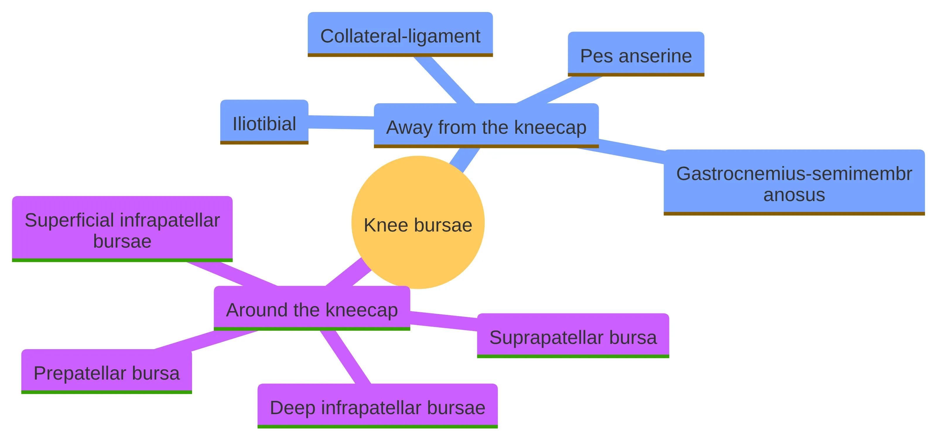
Knee bursae occur in several locations around the joint, so the place where your pain is concentrated helps define the question being assessed. Named areas include the prepatellar bursa in front of the kneecap, superficial and deep infrapatellar bursae below it, and the suprapatellar bursa. Other bursae sit away from the kneecap, including the pes anserine, iliotibial, collateral-ligament, and gastrocnemius-semimembranosus bursae.
That map is useful, but it is not a diagnosis. A front-of-knee complaint may raise a question about the prepatellar area. A complaint along the inner side of the knee may lead an evaluation toward the pes anserine area. A posterior or side-of-knee complaint creates a different set of structures to examine. Location narrows the discussion; it does not identify the source by itself.
Your description does not need to use anatomical terms. You can point to the most painful area, describe whether the pain is superficial or deep, and explain whether pressure on the area changes it. You can also note whether the discomfort stays in one spot or travels around the joint. Those details help a clinician decide which structures deserve attention.
MRI findings around a knee bursa need to be interpreted with the symptom pattern. Some bursae and recesses can contain a small amount of fluid without causing symptoms, and some are not visible on MRI when they contain little or no fluid. Findings that may support symptomatic bursitis include more prominent bursal distension, bursal-wall thickening, nearby soft-tissue or reactive marrow swelling, effects on a nerve or vessel, or signs of rupture.
MRI fluid therefore answers only part of the question. The meaningful question is whether the imaging abnormality sits in the same location as your symptoms and fits the way the problem began. A scan can identify a structure to discuss without proving that structure is the pain source.
Treat the anatomical map as a way to organize the evaluation, not as a way to diagnose yourself. If the painful area and the scan point to different structures, that mismatch is useful information. It is a reason to ask what else could account for the symptoms, not a reason to assume the scan is irrelevant.
Use the Symptom Story as a Clue, Not a Diagnosis
The symptom story gives a clinician context that an image cannot provide. Repeated minor trauma, such as frequent kneeling, is a common cause of superficial bursitis. Acute trauma or bleeding into the bursa, inflammatory disorders such as gout or rheumatoid arthritis, and infection are other possible causes. A direct injury and a gradually repeated pressure pattern can therefore lead to different questions during an evaluation.
Pain, tenderness, swelling, redness, and warmth may appear in a bursal problem, particularly when infection is involved, but those features overlap with other knee conditions. Pes anserine pain can resemble a stress fracture, which is one reason an assessment may include an X-ray. Symptoms alone can suggest a direction; they cannot confirm the tissue involved.
The timing of symptoms can also make the evaluation more precise. Tell the clinician whether the problem began after one event, followed repeated kneeling or another pressure pattern, appeared with skin changes, or returned after seeming to settle. Explain whether the discomfort is mainly present with pressure, walking, bending, or movement of the nearby joint. These details do not establish a diagnosis, but they help separate the questions that need answering.
A useful knee symptom history answers a few concrete questions:
- Where is the pain? Describe whether it is in front of the kneecap, below it, along the inner side, behind the knee, or elsewhere.
- What preceded it? Note repeated kneeling, a direct blow, another injury, or a change that placed pressure on the area.
- What does the skin look and feel like? Redness, warmth, swelling, a wound, or another skin break changes the assessment.
- How has the pattern changed? Tell the clinician whether pain and swelling appeared suddenly, built gradually, or keep returning.
- What other explanation needs to be excluded? A stress fracture, cellulitis, a septic joint, or another knee pain syndrome may require a different evaluation.
The point of these questions is not to assign yourself a label. The point is to give the examination a clear starting point and to identify whether prompt assessment is warranted. Write down the answers before an appointment if pain has made the sequence difficult to remember.
Check Whether “Bursitis” Explains the Knee Pain
The word “bursitis” describes inflammation of a bursa, but not every knee pain syndrome called bursitis actually involves a bursa. Pes anserine pain syndrome is one example of a condition that was formerly referred to as anserine bursitis. That distinction matters because a treatment aimed at an inflamed bursa may not address pain arising from another structure.
A second problem is that an abnormal scan can appear more definite than it is. Fluid in a bursa may be present without symptoms, while more substantial distension, wall changes, nearby swelling, nerve or vessel effects, or rupture may provide stronger imaging support. Even those features need to be compared with your location, onset, examination, and other possible causes.
The label can also hide different levels of urgency. Noninfectious irritation, a traumatic or hemorrhagic problem, a possible fracture, and suspected infection should not be handled as though they were the same condition. Before discussing a treatment, ask what clinical question the label is meant to answer and what finding would change the plan.
A clinician is trying to answer three separate questions when checking whether the label fits:
- Is a bursa located where the symptoms are centered? Anatomical location gives the suspected structure a reasonable starting point.
- Does the symptom pattern fit that structure? Repeated pressure, direct trauma, swelling, or tenderness can support the question, but none is conclusive alone.
- Could another condition explain the same presentation? Pes anserine pain may resemble a stress fracture, and infectious bursitis can resemble noninfectious inflammation or even a septic joint.
An evaluation that keeps these questions separate is more useful than an evaluation that treats the label as settled from the beginning. If the answers remain uncertain, the appropriate next step is clarification rather than a predetermined procedure.
Move Infection Warning Signs Out of the Wait-and-See Lane
Possible infection changes the next step for knee swelling or tenderness. Trauma or a break in the skin can be relevant to the history. These findings are not completely reliable for separating infectious from noninfectious bursitis, so a reassuring guess based on symptoms alone is not enough.
If you have that pattern, move out of the observation lane and seek prompt professional evaluation. Do not treat redness, warmth, or a skin break as proof that the problem is only mechanical irritation. A clinician may need to assess the bursa, the adjacent joint, and the surrounding skin and soft tissue.
Prompt evaluation does not mean that infection has been established. It means the possible consequence of missing an infectious process is significant enough that observation alone is not a sensible way to resolve the uncertainty. The warning signs should change the timing of the assessment, not be used to make a diagnosis at home.
Suspected infectious bursitis may require more than a visual inspection. Ultrasound can help distinguish bursitis from cellulitis. Blood testing and MRI may help assess infectious versus noninfectious causes. When infection is suspected, aspiration may be performed so bursal fluid can undergo laboratory analysis, including examination for organisms, crystals, glucose, and blood cells.
The exact testing sequence depends on the clinical situation. The practical boundary is clear: warning signs call for assessment rather than a self-directed attempt to identify the cause or choose a regenerative procedure. If a skin break, warmth, redness, swelling, or fever is part of your knee problem, make that information the first thing you report.
Understand What a Useful Evaluation Is Trying to Establish
A useful knee evaluation tries to connect your history, physical findings, and the most relevant test instead of treating any one piece as decisive. Superficial bursitis is usually assessed from the clinical presentation, with particular attention to possible infection. The examination may consider the size and consistency of swelling, whether fluid movement is present, skin changes, nearby cellulitis, lymph nodes, joint motion, and pain in the adjacent joint.
Your history supplies details that cannot be read from an image. Recent trauma, repeated pressure, a wound, inflammatory disease, and the sequence of swelling or pain can change the questions asked during the examination. The aim is to determine whether the suspected bursa fits the symptoms and whether another condition needs attention first.
An evaluation is therefore more than a search for a name. It should establish which structure is being considered, what makes that structure plausible, which warning signs affect urgency, and which uncertainty remains. That sequence lets the next decision follow the findings instead of forcing the findings to support a treatment that was selected in advance.
The evaluation may need to establish several boundaries before treatment is discussed:
- Bursa or another pain source: A syndrome labeled bursitis may not involve a bursa, and pes anserine pain can resemble a stress fracture.
- Infectious or noninfectious process: Symptoms and examination findings can overlap, so additional testing may be needed when infection is a concern.
- Soft-tissue question or bone question: An X-ray may be used to check for a broken bone when the presentation resembles a stress fracture; ultrasound or MRI may be used when more detail is needed.
- Symptomatic finding or incidental finding: MRI fluid does not automatically explain pain, so the scan must be matched to the clinical picture.
Those boundaries help keep the next decision proportional to what is actually known. They also give you useful questions to ask: What is the leading explanation? What competing explanation still needs to be excluded? What finding would change the urgency or treatment discussion?
Before leaving an evaluation, ask what has been considered, what remains uncertain, and what would change the plan. Ask which symptom change should prompt earlier follow-up, whether the available imaging is sufficient for the question, and what result would lead to a different pathway. These questions do not replace clinical advice; they make the plan easier to follow and reduce the chance that a provisional label is mistaken for a confirmed diagnosis.
Use Ultrasound and Prior Imaging to Answer a Specific Question
Diagnostic ultrasound is most useful when it answers a defined question about the painful area. Ultrasound may help distinguish bursitis from cellulitis, and it may help assess the extent of an injury when a clinician considers that information relevant. An ultrasound finding should still be interpreted alongside the location, skin findings, tenderness, joint motion, and symptom history.
An ultrasound image is not a substitute for an examination. It can add information about a suspected superficial structure, but it does not independently establish that the structure is responsible for every symptom or that a particular treatment is suitable. Ask what the ultrasound is intended to clarify before treating the image as the answer.
The value of an imaging test depends on the decision it informs. If the question is whether a superficial fluid collection is present, ultrasound may add useful detail. If the concern is a possible fracture or a deeper structural problem, another form of assessment may be relevant. The test should follow the clinical question rather than replace it.
Prior MRI and X-ray images can be useful when they answer the current clinical question. An X-ray may help check for a broken bone when pes anserine symptoms resemble a stress fracture. MRI may show fluid or other changes around a bursa, but fluid alone does not prove symptomatic bursitis. More prominent distension, wall thickening, nearby swelling, nerve or vessel effects, or rupture may add context, while symptoms remain part of the interpretation.
Bring prior reports and images to an evaluation when they are available. The useful question is not simply “What did the scan show?” It is “Does this finding match the place, timing, examination, and behavior of my pain?”
A prior scan is part of the record, not a final verdict. If your symptoms have changed since the image was taken, say so. If the report describes a finding in a different location from your pain, point out that difference. The interpretation should account for the current clinical picture.
Choose the Least-Assumptive Care Pathway First
A sensible care pathway depends on what remains uncertain. Use the following branches as a way to organize the next conversation, not as a personal treatment prescription.
If the pattern is clearly acute, traumatic, or hemorrhagic and infection warning signs are absent: cited guidance describes conservative measures such as ice, elevation, rest, and analgesics. Discuss which measures fit your situation with a qualified clinician, and do not treat a suspected label as confirmed merely because the symptoms seem familiar.
If the pain is persistent, recurrent, or difficult to localize: arrange an evaluation. The reason is not that every persistent symptom needs an advanced procedure; it is that the diagnosis, imaging interpretation, and next step remain unclear. A history and examination can determine whether targeted ultrasound, an X-ray, MRI, or another assessment is relevant.
If the symptoms include redness, warmth, swelling, a skin break, or fever: move out of the wait-and-see lane and seek prompt professional evaluation. Symptoms alone cannot reliably distinguish septic from noninfectious bursitis, and suspected infection may require aspiration and fluid analysis.
If the pattern resembles a stress fracture: make the bone question part of the evaluation. Pes anserine pain can resemble a stress fracture, and an X-ray may be used to check for a broken bone. Do not let a presumed bursa label close that question.
Each branch ends with a different action because each leaves a different level of uncertainty. The absence of warning signs may make a conservative discussion reasonable in a clearly acute pattern. Persistence, mismatch, or concern about infection or fracture changes the action to evaluation.
Conservative care is a pathway, not a promise that the first label is correct. A response or lack of response can change the next discussion, but neither one proves which structure is involved. The same principle applies to imaging: a scan can add detail without deciding the source of pain.
Start with the least-assumptive pathway that fits the known facts. Escalate the evaluation when the facts do not fit together, when symptoms continue, or when warning signs change the level of concern. Keep a short record of the pain location, swelling, skin changes, triggers, and changes over time so that the next discussion begins with a clear account rather than a vague treatment request.
Use a change in the pattern as a checkpoint rather than waiting for the original explanation to prove itself. If pain that was easy to localize becomes diffuse, swelling increases, or the skin develops new changes, tell a clinician what changed and when. If symptoms settle but return with the same pressure or activity, record that recurrence instead of assuming it confirms bursitis. These observations do not diagnose the cause; they show whether the working explanation still fits. They can also help determine whether the next question concerns a superficial bursa, a deeper structure, a possible bone problem, or a process involving the skin or joint. A useful follow-up conversation compares the current pattern with the starting pattern, the response to conservative measures, and any available imaging. That comparison keeps care proportional: observation remains reasonable only while the known facts support it, and assessment becomes more important when the pattern no longer matches.
Keep Regenerative Options Downstream of the Assessment
Regenerative treatment should come after the cause and urgency of the knee problem have been assessed. Infection, a possible fracture, a pain syndrome that does not involve a bursa, and an incidental MRI finding create different clinical questions. None should be bypassed by choosing a procedure from the word “bursitis” alone.
RegenCore Method provides non-surgical treatments including PRP, bone marrow concentrate, stem cell therapy, shockwave, EMTT, and spinal disc care. That list describes available treatment categories; it does not establish that every option applies to a knee bursa problem, every symptom pattern, or every person. Suitability still depends on the assessment and the condition being addressed.
The practical order is simple: establish what may be causing the pain, address any urgent concern, interpret imaging in context, and then discuss treatment options that relate to the findings. This order protects the assessment from being shaped by the availability of a procedure. It also gives you a clearer basis for deciding whether a non-surgical option deserves further discussion.
A focused discussion about a non-surgical option should answer practical questions before a procedure is considered:
- What structure or condition is the treatment intended to address?
- What findings support that interpretation?
- Has infection been considered when redness, warmth, swelling, skin breaks, or fever are present?
- Has a possible stress fracture or another non-bursal cause been considered when the symptom pattern calls for it?
- What would conservative care or further evaluation establish before an intervention?
PRP, bone marrow concentrate, shockwave, and other options should remain downstream of those questions. An option can be available without being the right answer to the problem under discussion. If the cause of pain is still uncertain, the next useful step is to clarify the diagnosis and suitability question rather than compare procedures in the abstract.
What a RegenCore Method Evaluation Can Add
A RegenCore Method evaluation includes a thorough history, hands-on orthopedic exam, diagnostic ultrasound, review of prior MRI or X-ray imaging, and a personalized treatment and recovery plan. That sequence gives you a place to connect the symptom story with the examination and available imaging instead of discussing a treatment category in isolation.
Bring a concise account of when the pain began, where it is centered, whether repeated pressure or trauma preceded it, how swelling or skin changes have changed, and what prior imaging is available. Those details help keep the conversation focused on the structure and question that need clarification.
You can prepare for the evaluation by organizing:
- the date or circumstances when symptoms began;
- the location and character of the pain;
- any repeated kneeling, direct injury, wound, redness, warmth, or swelling;
- changes in symptoms since prior imaging; and
- the questions you want answered before considering treatment.
This preparation does not determine the outcome. It makes the clinical discussion more specific.
RegenCore Method can guide you across the full spectrum of care and reserve surgery for when it is truly the right answer. That approach does not turn an evaluation into a promise of regenerative treatment. It keeps the order intact: clarify the cause, assess the relevant findings, discuss suitable paths, and then decide what deserves consideration.
If your symptoms are unclear, persistent, or difficult to match with an imaging finding, an evaluation can turn that uncertainty into a specific clinical discussion. If infection warning signs are present, seek prompt professional assessment rather than waiting for a regenerative-treatment conversation to resolve the issue. The evaluation is a place to discuss what the findings support, not a commitment to a particular intervention.
Book the Conversation When the Next Step Is Still Unclear
The right next step for suspected knee bursitis depends on the pattern, not the label alone. Use the symptom location and trigger to frame the question, check for redness, warmth, swelling, skin breaks, or fever, and treat MRI fluid as a finding that still needs clinical context. Acute traumatic or hemorrhagic patterns may lead to a conservative-care discussion, while persistent, unclear, or concerning symptoms deserve evaluation. A RegenCore Method evaluation includes history, orthopedic examination, diagnostic ultrasound, prior imaging review, and a personalized treatment and recovery plan.
A qualified professional must decide individual suitability, including whether a suspected bursa problem is present and which care pathway fits your findings. If the cause of your knee pain remains unclear, bring your symptom timeline and prior imaging to a focused evaluation so the next decision can follow the actual clinical picture.
Next step
Have a question for RegenCore?
Concierge regenerative & orthobiologic orthopedics in San Francisco and Palo Alto. Research-backed, non-surgical treatment — PRP, bone marrow concentrate (BMAC), stem cell therapy, shockwave, EMTT and spinal disc care — to help the body heal itself.
Get in touch