
Understanding Musculoskeletal Conditions
Understanding X-Rays of Arthritis: Diagnosis, Limitations, and Alternatives

Introduction
For millions living with arthritis, the path to understanding their condition can feel overwhelming and isolating. While X-rays are often the first step in diagnosing arthritis, many patients find themselves feeling lost and overwhelmed, unsure of where to turn for answers. These imaging techniques come with limitations that can leave patients frustrated and in the dark about their health.
But what if there were better ways to uncover the truth about their health? This article will explore how X-rays fit into the bigger picture of arthritis diagnosis, discuss their limitations, and shine a light on advanced imaging options that could empower patients to better understand and manage their condition.
Define Arthritis and Its Types
Imagine waking up every day with pain that limits your ability to enjoy life, just like the 32 million Americans suffering from osteoarthritis. Arthritis is more than just a medical term; it encompasses over 100 conditions that can turn everyday activities into painful struggles, leaving many feeling isolated and frustrated. Among these, osteoarthritis (OA) stands out as the most common, affecting millions and often leading to a painful journey of reduced mobility and frustration. This degenerative condition occurs when the cartilage that cushions the connections between bones deteriorates over time, resulting in pain that can feel overwhelming.
Rheumatoid arthritis (RA) adds another layer of complexity, as it’s an autoimmune condition where the immune system mistakenly attacks the synovium, the lining of the joints. This can lead to inflammation and pain, disproportionately affecting women, who are two to three times more likely to be diagnosed than men.
Then there’s psoriatic arthritis, which is linked to the skin condition psoriasis. This type can cause swelling, pain, and stiffness in the joints, impacting overall health and quality of life. Gout, characterized by sudden and severe pain, often strikes unexpectedly, particularly in the big toe, leaving individuals in agony due to the buildup of uric acid crystals.
Ankylosing spondylitis primarily affects the spine, leading to severe inflammation and pain in the vertebrae, which can significantly hinder mobility. Understanding these types of arthritis is crucial, not just for diagnosis, but for recognizing the struggles faced by those living with these conditions every day.
Recent studies suggest that by 2040, around 78.4 million adults in the U.S. will be diagnosed with joint inflammation, with many facing activity limitations. This highlights the ongoing need for effective management strategies and innovative treatments in the field of regenerative medicine. With the right support and innovative treatments, a life free from the constraints of arthritis is within reach.

Explain How X-Rays Diagnose Arthritis
Imagine waking up each day with joint pain that makes even the simplest tasks feel daunting. Radiographs play a vital role in understanding the state of your bones and joints, helping to identify arthritis and its impact on your life.
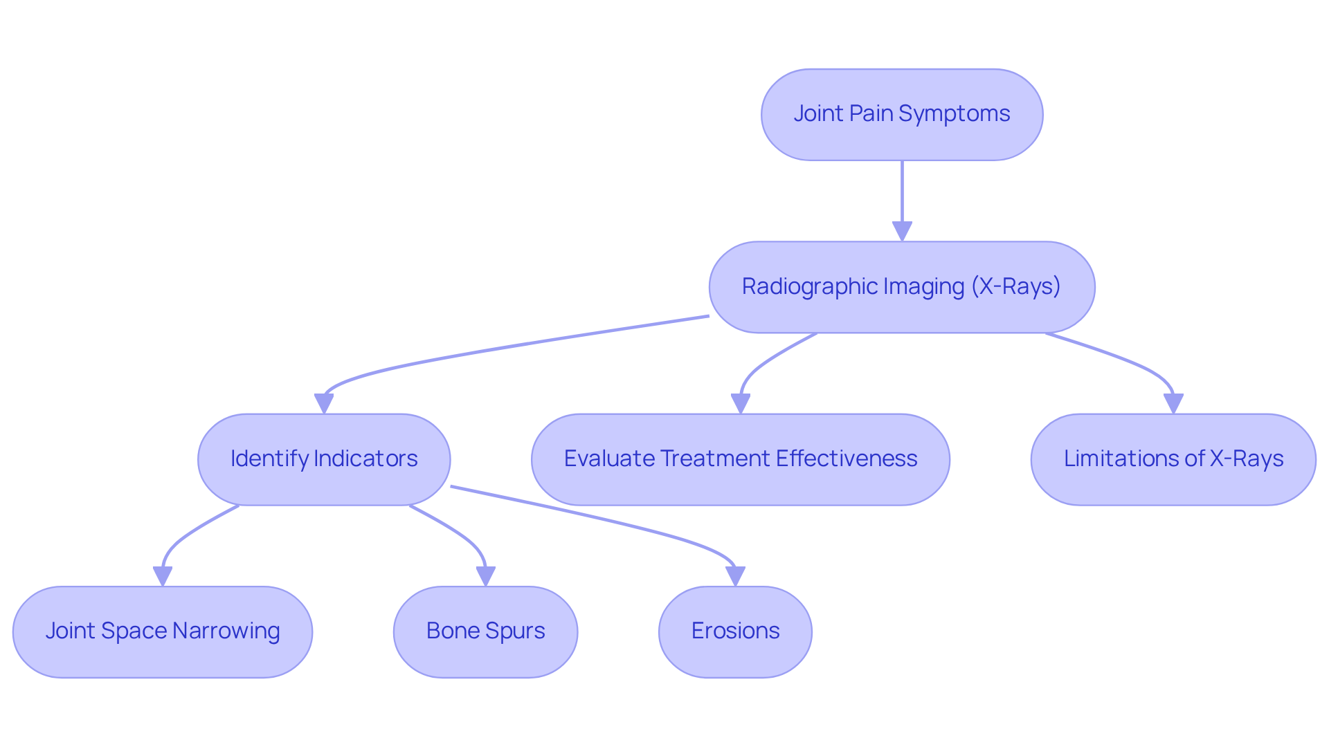
When you share your struggles with joint discomfort, radiographic imaging often becomes the first step in uncovering the truth behind your pain. These images provide a clear view of joint structures and any visible alterations, allowing physicians to evaluate rheumatoid joint inflammation (RA) by examining both hands and feet.
X-rays of arthritis can highlight several critical indicators, including joint space narrowing, which reflects cartilage loss and is a key sign of osteoarthritis. You might also see bone spurs, those bony projections that often develop along joint margins, particularly in osteoarthritis cases. In inflammatory forms like rheumatoid joint disease (RA), radiographic imaging can show erosions on the bone surface, indicating disease progression. It’s significant to note that radiographs of the hands and feet may reveal RA erosion just 1 to 2 years after the onset of the disease.
As time goes on, radiographic imaging remains essential in observing the advancement of joint inflammation. These images allow healthcare providers to evaluate the effectiveness of treatment plans by comparing changes in joint structure. They are often the initial diagnostic tests employed to identify indications of joint inflammation.
Yet, it’s important to remember that while radiographs are helpful, they have their limitations. They may not effectively capture early-stage joint inflammation or soft tissue changes, which is why they are often supplemented with advanced imaging techniques like MRI or ultrasound. Research shows that MRI can identify bone erosions up to three years prior to their visibility on radiographs, highlighting the significance of a thorough diagnostic strategy. As mentioned by the Raleigh Bone and Joint Surgery Clinic, “Radiographic imaging is a dependable method to recognize moderate to advanced joint inflammation, but less reliable in spotting early indicators of the condition.”
By combining various imaging techniques, we can ensure that no patient has to face the challenges of arthritis alone, receiving the timely care they deserve.
Identify Limitations of X-Ray Imaging for Arthritis
Imagine living with joint pain, seeking answers, only to be told that your X-rays look normal, leaving you feeling unheard and frustrated. While X-rays of arthritis serve as a common diagnostic tool, they present several limitations in diagnosing the condition that can leave patients feeling lost.
Early detection can be particularly challenging. Radiographic imaging often fails to reveal abnormalities in the initial stages of arthritis, especially in inflammatory types where soft tissue inflammation is predominant. For instance, a study found that osteophytes were only seen 49.7% of the time on X-ray despite being present on clinical exams. This highlights the importance of a comprehensive evaluation, which is a cornerstone of the RegenCore Method‘s approach.
Moreover, X-rays focus primarily on bony structures, leaving out crucial information about cartilage, ligaments, or synovial tissue. These elements are essential for a complete assessment of joint conditions. MRI can provide sharper images of osteoarthritis when X-ray results fall short, identifying cartilage defects that traditional imaging may overlook. At RegenCore, we understand how vital it is to see the whole picture, so we combine advanced imaging techniques with compassionate care to ensure you feel supported every step of the way.
There’s also a disconnect between the severity of arthritis symptoms and X-rays of arthritis findings. Some patients endure significant pain despite normal X-ray results, while others may exhibit severe X-ray findings with minimal symptoms. Dr. Jolanda Cibere has stated that routine use of plain radiography in osteoarthritis patients is not indicated, highlighting this issue. RegenCore addresses this variability through personalized treatment plans that consider your unique pain experiences and functional limitations.
Additionally, the interpretation of X-ray images can vary, leading to false positives or negatives that may influence treatment decisions. The Kellgren-Lawrence system for grading osteoarthritic structures suffers from ambiguity, particularly between grades 0 and 1, complicating evaluations. Our team at RegenCore emphasizes a detailed orthopedic evaluation to mitigate these risks and ensure accurate diagnoses.
Given these constraints, X-rays are often used alongside other scanning techniques, such as MRI or ultrasound, to obtain a more thorough evaluation of limb conditions. Furthermore, weight-bearing positions are essential for precise X-ray assessment of space narrowing, particularly in hips and knees. At RegenCore, we combine these visual insights with our innovative regenerative therapies, including Bone Marrow Aspirate Concentrate (BMAC) and Platelet-Rich Plasma (PRP), to enhance healing and promote tissue repair.
If you’re experiencing joint pain or arthritis symptoms, don’t let uncertainty dictate your journey; reach out to us at RegenCore, where we prioritize your unique experience and healing path. Contact us at 650-328-4411 or visit us at 2248 Park Blvd., Palo Alto, CA 94306 to discuss your condition and explore personalized treatment options.

Explore Alternative Imaging Techniques for Arthritis Diagnosis
For those living with arthritis, the journey to understanding their condition can often feel overwhelming and frustrating. Many patients struggle with the uncertainty of their diagnosis, feeling as if their pain is invisible to the world. This is where advanced imaging techniques come into play, offering a glimmer of hope.
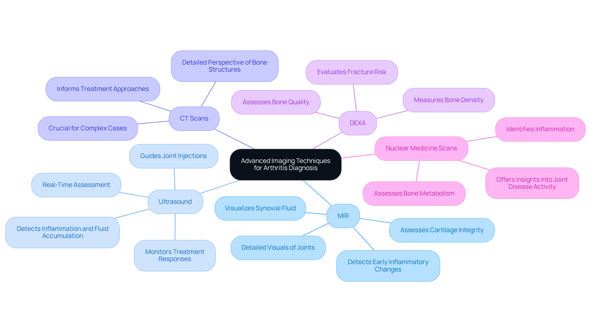
- Magnetic Resonance Imaging (MRI) can be a game-changer. Imagine the relief of seeing clear images of your joints, revealing the hidden struggles of arthritis that you’ve felt for so long. MRI provides detailed visuals of both bone and soft tissues, making it particularly useful for detecting early inflammatory changes and assessing cartilage integrity. It can visualize synovial fluid and soft tissue structures that traditional imaging cannot, giving patients a clearer picture of their condition.
- Then there’s Ultrasound. This technique uses sound waves to create images of soft tissues, helping to detect inflammation, fluid accumulation, and synovitis in joints. Picture a doctor guiding an injection with precision, all thanks to ultrasound. It’s especially beneficial for monitoring treatment responses, allowing patients to see their progress in real-time.
- Computed Tomography (CT) scans offer another layer of insight. They provide a more detailed perspective of bone structures than traditional imaging, which can be crucial in complex cases where accurate anatomical details are required. For patients, this means a more informed approach to their treatment.
- Dual-Energy X-ray Absorptiometry (DEXA) is primarily known for measuring bone density, but it can also shed light on bone quality and help assess the risk of fractures in arthritic patients. Understanding these risks can empower patients to take proactive steps in their care.
- Lastly, Nuclear Medicine Scans can identify inflammation and changes in bone metabolism, offering insights into the activity of joint disease that may not be visible on X-rays. This knowledge can be invaluable for patients seeking clarity in their treatment journey.
By embracing these advanced imaging techniques, patients can find renewed hope and clarity in their treatment journey, paving the way for a better quality of life.
Conclusion
Navigating the complexities of arthritis can feel overwhelming, leaving many patients unsure of their next steps. Understanding arthritis and its nuances is essential for anyone facing the challenges of joint pain. While X-rays play a critical role in diagnosing arthritis, they often fall short in detecting early-stage issues and soft tissue problems. This highlights the importance of exploring advanced imaging techniques like MRI and ultrasound, which can provide a clearer picture of joint health.
Key insights reveal that while X-rays can show significant changes in joint structure, they may not capture the full extent of arthritis. This underscores the need for personalized treatment plans that consider each individual’s experiences and symptoms, ensuring that patients receive the most effective care tailored to their needs.
But remember, you’re not alone in this journey; taking proactive steps can lead to a brighter future. Embracing advanced imaging techniques and seeking comprehensive evaluations empowers patients to take control of their health. For those experiencing joint pain or arthritis symptoms, reaching out for support and exploring innovative treatment options, such as those offered by the RegenCore Method, can transform your journey into one of empowerment and hope. Contact RegenCore at 650-328-4411 or visit 2248 Park Blvd., Palo Alto, CA 94306 to discuss personalized solutions for your arthritis journey.
Frequently Asked Questions
What is arthritis?
Arthritis is a term that encompasses over 100 conditions that cause pain and inflammation in the joints, significantly impacting daily activities and quality of life.
What are the most common types of arthritis?
The most common types of arthritis include osteoarthritis (OA), rheumatoid arthritis (RA), psoriatic arthritis, gout, and ankylosing spondylitis.
What is osteoarthritis and how does it affect individuals?
Osteoarthritis is the most common form of arthritis, characterized by the deterioration of cartilage that cushions the joints, leading to pain, reduced mobility, and frustration for those affected.
How does rheumatoid arthritis differ from osteoarthritis?
Rheumatoid arthritis is an autoimmune condition where the immune system attacks the synovium, causing inflammation and pain in the joints, and it disproportionately affects women.
What is psoriatic arthritis?
Psoriatic arthritis is linked to the skin condition psoriasis and can cause swelling, pain, and stiffness in the joints, negatively impacting overall health and quality of life.
What are the symptoms of gout?
Gout is characterized by sudden and severe pain, often in the big toe, due to the buildup of uric acid crystals, causing intense agony for those affected.
What is ankylosing spondylitis?
Ankylosing spondylitis primarily affects the spine, leading to severe inflammation and pain in the vertebrae, which can significantly hinder mobility.
What is the projected future of arthritis diagnoses in the U.S.?
Recent studies suggest that by 2040, approximately 78.4 million adults in the U.S. will be diagnosed with joint inflammation, highlighting the need for effective management strategies and innovative treatments.
List of Sources
- Define Arthritis and Its Types
- New Research Brings Joint Repair Closer for Millions With Osteoarthritis | Duke Department of Orthopaedic Surgery (https://ortho.duke.edu/news/new-research-brings-joint-repair-closer-millions-osteoarthritis)
- Arthritis-associated activity limitations reported in more than 24 million US adults (https://healio.com/news/rheumatology/20260126/arthritisassociated-activity-limitations-reported-in-more-than-24-million-us-adults)
- The Lancet: New study reveals the most common form of arthritis, osteoarthritis, affects 15% of the global population over the age of 30 (https://healthdata.org/news-events/newsroom/news-releases/lancet-new-study-reveals-most-common-form-arthritis)
- Rheumatoid arthritis (https://who.int/news-room/fact-sheets/detail/rheumatoid-arthritis)
- Home – Rheumatology Advisor (https://rheumatologyadvisor.com/news/scientists-test-new-ways-to-regrow-joints-damaged-by-arthritis)
- Explain How X-Rays Diagnose Arthritis
- Rheumatoid Arthritis X-Ray: Diagnosis and Evaluation (https://healthcentral.com/article/can-you-see-rheumatoid-arthritis-on-x-rays)
- Do X-Rays Show Arthritis & What Do Your Scan Results Mean? (https://raleighboneandjoint.com/news/do-x-rays-show-arthritis)
- Use of Imaging Scans in the Detection and Monitoring of RA (https://arthritis.org/health-wellness/treatment/treatment-plan/disease-management/imaging-for-rheumatoid-arthritis)
- What Do You See on X-Rays If You Have Rheumatoid Arthritis? (https://healthline.com/health/rheumatoid-arthritis/x-rays)
- How Does Diagnostic Imaging Improve Arthritis Treatment Plans? (https://glmi.com/blog/how-does-diagnostic-imaging-improve-arthritis-treatment-plans)
- Identify Limitations of X-Ray Imaging for Arthritis
- Plain x-rays not recommended for confirming osteoarthritis (https://auntminnie.com/clinical-news/article/15576834/plain-x-rays-not-recommended-for-confirming-osteoarthritis)
- How X-Rays Help Diagnose and Monitor Rheumatoid Arthritis – IPMC – Independent Physicians Medical Center (https://ipmcmed.com/how-x-rays-help-diagnose-and-monitor-rheumatoid-arthritis)
- Will X Rays Show Arthritis? Diagnosis Insights (https://allhealthdtc.com/will-x-rays-show-arthritis-diagnosis-insights)
- Do X-Rays Show Arthritis & What Do Your Scan Results Mean? (https://raleighboneandjoint.com/news/do-x-rays-show-arthritis)
- Explore Alternative Imaging Techniques for Arthritis Diagnosis
- Imaging in arthritis: quantifying effects of therapeutic intervention using MRI and molecular imaging (https://smw.ch/index.php/smw/article/download/1631/2145?inline=1)
- Ultrasound Eases Arthritis Diagnosis (https://cuimc.columbia.edu/news/ultrasound-eases-arthritis-diagnosis)
- AxSpA Imaging and Treatment Updates (https://aiarthritis.org/axspa-imaging-and-treatment-updates)
- Advances in Imaging of Rheumatoid Arthritis | WCG (https://wcgclinical.com/insights/advances-in-imaging-of-rheumatoid-arthritis)
- Advanced Imaging Techniques to Transform Arthritis Diagnosis (https://emjreviews.com/rheumatology/news/advanced-imaging-techniques-to-transform-arthritis-diagnosis)
Next step
Have a question for RegenCore?
Concierge regenerative & orthobiologic orthopedics in San Francisco and Palo Alto. Research-backed, non-surgical treatment — PRP, bone marrow concentrate (BMAC), stem cell therapy, shockwave, EMTT and spinal disc care — to help the body heal itself.
Get in touch