
back of knee hurts
Back of Knee Pain: A Decision Guide for Choosing Your Next Step

Start with a posterior-knee symptom map
Pain behind your knee is a symptom location, not a diagnosis. The same area can hurt after an injury, alongside a problem involving muscle or tendon structures, or with a less common condition. The range of possible causes makes the posterior location a starting point for a careful history and examination, not a diagnosis.
A useful next step is to move through three questions in order:
- How does the posterior pattern behave? Note when it began, where it is located, how it feels, whether there was trauma, and how long it has lasted.
- Which accompanying findings need to be reported? Note swelling, instability, changes in motion or weight-bearing, warmth, redness, fever, or broader symptoms.
- What remains unknown? Use the history and examination to define the question before imaging or treatment is discussed.
This approach keeps you from assigning a cause to a symptom before its context is understood. It also keeps treatment choices downstream of the information that makes them relevant.
An opening symptom map works best when each observation stays in its proper role. Onset and duration describe the timeline; location and quality describe the experience; a fall, twist, impact, or activity change supplies context. Swelling, mechanical symptoms, broader symptoms, and relevant medical or surgical history add information for the clinical history. The value of the list is not that it produces a label. Its value is that it preserves details that can be examined together.
Function adds a separate dimension to the account. Weight-bearing ability, range of movement, instability, and changes to ordinary activity describe what the leg can do now. Those details are different from naming the tissue involved. They can show how the symptom affects daily life while leaving the cause open for evaluation. A symptom that is intermittent, recurring, or persistent also has a timeline that belongs in the account rather than being reduced to a single pain description.
An account can separate the initial event from the current state. A symptom may have a clear precipitating injury, or it may have begun without a remembered event; either fact belongs in the history. A return after temporary improvement is also timeline detail. Recording these changes helps keep one moment from standing in for the whole story and gives a clinician more context for the examination.
The findings around the knee matter alongside the location. Swelling, restricted movement, instability, mechanical symptoms, fever, warmth, redness, and symptoms beyond the knee are not interchangeable observations. They belong in separate parts of the description because they can change which questions need attention first. The warning-sign discussion addresses urgency as its own question, rather than using the posterior location to assign a cause.
The evaluation question is also distinct from the symptom description. A clinical history can organize onset, location, duration, quality, trauma, associated findings, and relevant medical or surgical history. The examination may then include inspection, palpation, range-of-motion and strength assessment, neurovascular testing, and focused tests. Together, these steps help define what remains uncertain and whether further evaluation is needed.
Imaging belongs to that same sequence. The cited initial-evaluation guidance discusses radiographs for chronic knee pain lasting more than six weeks or for acute traumatic pain that meets specific criteria. That does not turn duration into a diagnosis or make one study appropriate for every posterior-knee symptom. It means that the imaging question is connected to the history and examination findings that precede it.
Treatment discussion comes later for the same reason. The fact that pain is located behind the knee does not by itself establish that PRP, BMAC, shockwave therapy, or another option is relevant. A nonsurgical conversation has to remain connected to the clinical question, the information already available, and the uncertainty that still needs to be addressed. This keeps the article focused on choosing the next step rather than choosing a procedure from a symptom location.
The location is still useful. It tells you where to begin describing the problem, but not how serious it is or which structure is involved. Your next decision should therefore depend on the combination of location, onset, injury history, swelling, movement, weight-bearing ability, and broader symptoms.
For example, pain that began after a twist raises a different evaluation question from pain that appeared gradually without a clear event. Pain with fever or a warm, swollen joint belongs on a more urgent pathway than mild pain without those findings. These distinctions do not diagnose you. They help you choose the appropriate level of attention.
Sort the possibilities by tissue and context
Pain behind your knee can come from more than one category, and the pattern around the pain matters as much as its location. A review of posterior knee pain describes a broad range of possible causes, including common injuries involving musculotendinous structures and less common conditions. That range is why a symptom label should remain a starting point rather than a conclusion.
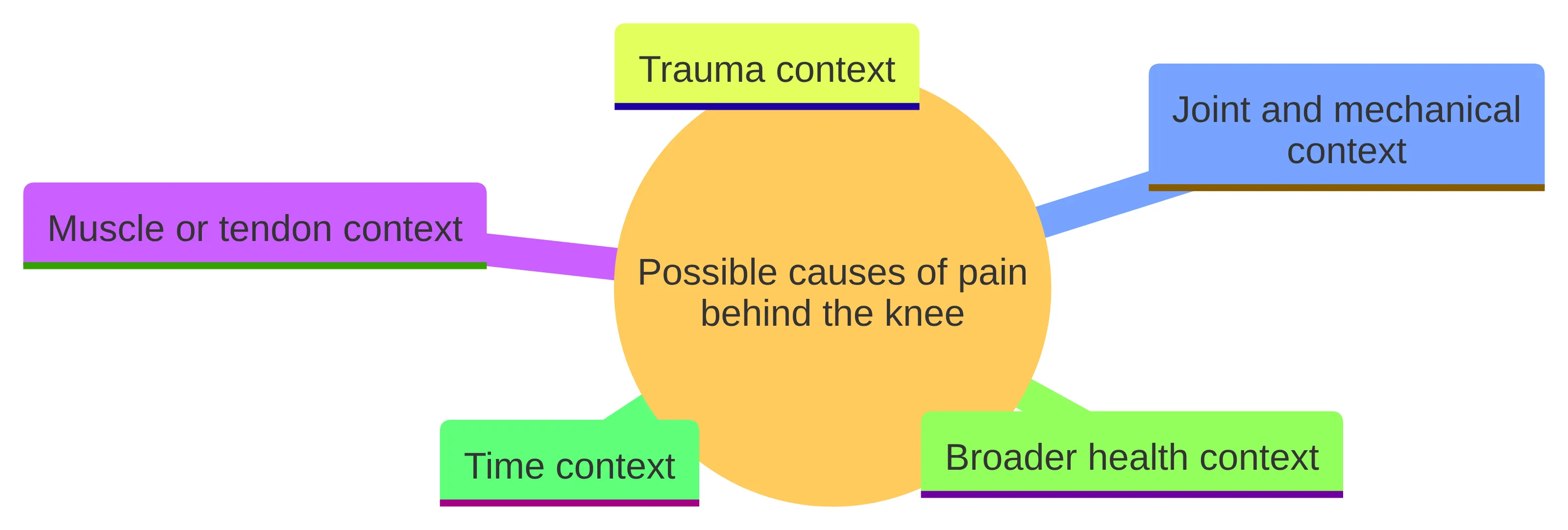
A clinician will usually sort the story by tissue and context rather than by location alone:
- Muscle or tendon context: Pain that follows a clear strain or a particular activity may raise questions about structures around the back of the knee. The timing, trigger, and effect on movement still need to be considered together.
- Joint and mechanical context: Swelling, restricted motion, or other mechanical symptoms can change the questions asked during the examination. These clues do not identify a diagnosis by themselves.
- Trauma context: A fall, twist, direct impact, or sudden change in function can make the injury mechanism central to the next decision.
- Broader health context: Fever, redness, warmth, or symptoms outside the knee can make a routine musculoskeletal explanation less suitable as a working assumption.
- Time context: The difference between pain that started today, pain that keeps returning, and pain that has lasted more than six weeks can affect how an evaluation proceeds.
Your description becomes more useful when it connects the symptom to an event, activity, duration, swelling, mechanical symptoms, or broader symptoms. No single detail should be used to declare the cause.
Before an appointment, organize the story around the questions that separate these categories:
- Did the pain begin suddenly or gradually?
- Was there a fall, twist, impact, new workout, or other change before it started?
- Is the pain constant, intermittent, or tied to a particular movement?
- Have you noticed swelling, catching, locking, instability, weakness, or a reduced range of motion?
- Can you bear weight and walk as usual?
- Has the symptom improved, stayed the same, worsened, or returned after temporary improvement?
- Do fever, redness, warmth, or other symptoms accompany the knee problem?
This information does not replace an examination. It gives the examination a clearer starting point and helps prevent the discussion from narrowing too quickly to a single presumed cause.
Screen the posterior pattern for findings that change urgency
For a posterior-knee symptom, accompanying findings determine urgency before any treatment category is compared. Seek prompt medical evaluation if severe knee pain occurs with swelling and instability or an inability to bear weight after acute trauma. Seek prompt evaluation as well for fever, a red or swollen joint, warmth, or limited range of motion.
A warm, swollen, red knee can signal a potentially serious problem such as infection. Do not treat that pattern as ordinary soreness or wait for a treatment comparison to resolve it.
Use the following distinction:
- After an injury: severe pain combined with swelling, instability, or inability to bear weight calls for prompt medical attention.
- With possible joint inflammation or infection: fever, warmth, redness, swelling, and limited motion call for prompt medical attention.
- Without those warning signs: document the pattern and arrange a suitable assessment when the symptom persists, returns, limits activity, or remains unclear.
These signs do not tell you the final diagnosis. They tell you that the next step should focus on medical evaluation rather than self-selecting a treatment.
Urgency is about the combination of findings, not the word used to describe the pain. “Sore,” “tight,” or “aching” does not by itself establish that a symptom is minor. A less dramatic description can still deserve evaluation if you cannot bear weight, the joint is becoming swollen and difficult to move, or fever and redness are present.
If a warning sign is present, do not spend the next step comparing PRP, BMAC, shockwave therapy, or other options. First obtain the medical attention appropriate to the warning pattern. Treatment selection belongs after the immediate concern has been assessed.
Read the posterior pattern across context, companions, and function
Read pain behind the knee as a pattern across three dimensions: what preceded it, what accompanies it, and what the leg can do now. Note the onset and trigger first; then record changes in swelling, movement, weight-bearing, or ordinary activity. This keeps the posterior location central without treating it as a cause.
Use those dimensions to choose the question for the next step:
- Evaluation question: If the pattern is not urgent but pain persists, returns, limits activity, or remains unclear, what history and examination findings need to be established?
- Information question: If the symptom is mild, record changes in onset, trigger, duration, function, and associated findings. That record can make a later clinical discussion more focused.
Time remains one part of the posterior-knee account. The cited initial-evaluation guidance discusses radiographs for chronic knee pain lasting more than six weeks or for acute traumatic pain that meets specific criteria. That guidance organizes a clinical question; it does not diagnose the cause from duration alone.
Function gives you a more useful checkpoint than a pain score alone. Record whether you can bear weight, whether the knee moves as usual, and which ordinary activities have changed. Also note whether swelling, instability, or a mechanical symptom appeared with the pain.
Time adds another layer. A symptom that began after a known injury may need an injury-focused history and examination. Pain that persists beyond six weeks enters the chronic-pain framework described in the initial-evaluation guidance. Recurrent or unclear pain deserves attention even when it is not severe, because the missing information may be the reason you cannot decide what to do next.
The practical boundary is simple: use symptom duration to organize an evaluation, not to diagnose yourself or to assume that a particular imaging study or treatment is automatically required.
Keep interim decisions cautious and information-focused
Monitoring pain behind your knee should mean collecting decision-changing information, not trying to name the condition from home. A useful symptom record includes:
- when the pain began and whether it started suddenly or gradually;
- the exact location, quality, and pattern of the pain;
- any fall, twist, impact, or activity change before it began;
- swelling, limited motion, instability, or other mechanical symptoms;
- fever, redness, warmth, or symptoms beyond the knee;
- whether you can bear weight and which activities now cause difficulty; and
- relevant medical, surgical, or previous knee history.
This list follows the elements typically considered in a knee-pain history. It gives you a way to describe change without turning a symptom pattern into a diagnosis.
Keep prior MRI or X-ray images and reports available if you arrange an assessment. Reviewing prior imaging can be part of the process, but an older scan does not automatically answer a new question. The current history and examination still determine what needs to be clarified.
A short daily note can make this information easier to use. Record the date, what you were doing when the symptom appeared or changed, the location and quality of the pain, swelling or motion changes, and whether weight-bearing or a usual activity became harder. Add any relevant medical or surgical history and the names of prior imaging studies if you have them.
Bring the record to an appointment or use it when deciding whether the symptom is persistent, recurring, or activity-limiting. Its purpose is practical: it helps you communicate the pattern and gives the clinician a clearer timeline. It is not a home diagnostic test.
Do not let a monitoring plan override a warning sign. Fever, a warm or red swollen knee, severe pain after an injury, instability, inability to bear weight, or limited motion changes the next step to prompt medical evaluation.
Turn the posterior location into an evaluation question
An evaluation should turn the posterior location into a more specific clinical question. The history can establish when the pain began, where it is centered, how it feels, whether trauma preceded it, how long it has lasted, and whether swelling, mechanical or broader symptoms, or relevant medical or surgical history changes the picture.
The physical examination may include:
- looking at and feeling the knee;
- checking range of motion and strength;
- assessing nerve and circulation function; and
- performing focused examination tests.
These steps help determine whether the pattern is more consistent with an injury question, a joint question, a broader medical concern, or a need for imaging. The examination also supplies context for interpreting a scan rather than treating the scan as the answer by itself.
For pain behind the knee, the useful outcome is not simply a label. It is a clearer account of what is known, what remains uncertain, what needs attention first, and which treatment discussions relate to the unanswered question.
You can make the appointment more productive by bringing the symptom timeline, a list of prior injuries or procedures, relevant medical history, and prior imaging reports or images. Be ready to describe what you can and cannot do with the leg, not only how the pain feels.
Useful questions include:
- What part of my history or examination matters most here?
- Is the main question about the joint, surrounding soft tissue, the injury mechanism, or something broader?
- What finding would make imaging useful?
- What remains uncertain after the examination?
- Which next step follows from the findings rather than from the symptom location alone?
An evaluation does not have to produce every answer at once to be useful. It should clarify the immediate concern and identify the information needed for the next decision.
Make imaging answer the posterior question
For pain behind the knee, imaging should answer a question left by the history and examination rather than simply label the location. The question may involve an acute injury, a persistent symptom pattern, or a finding that the initial assessment cannot explain.
The cited initial-evaluation guidance discusses radiographs for chronic knee pain lasting more than six weeks or for acute traumatic pain meeting specific criteria. That guidance keeps the posterior location in context: duration, injury history, and examination findings determine what information a study is meant to provide.
An imaging discussion should therefore answer four practical questions:
- What finding is the scan intended to investigate?
- What part of the examination or injury history created that question?
- What would a normal result change about the next step?
- What would an abnormal result change?
Those questions help keep imaging connected to your care instead of turning it into a search for any abnormality.
Prior imaging can be useful, but its value depends on the question being asked now. Tell the clinician when the earlier scan was performed, what had happened before it, and whether the current symptoms are the same or different. A prior MRI or X-ray may provide context without settling a new symptom pattern.
For an acute injury, the examination findings and mechanism help determine whether radiographs are an appropriate initial study. For persistent pain, the duration and overall history help place imaging in the broader evaluation. A scan should follow that reasoning, not replace it.
If imaging is proposed, ask what decision it is meant to inform. Knowing that purpose helps you understand why a particular study is being considered and what the next step could be if the result is normal or abnormal.
Resolve the imaging guidance through the injury pattern and examination
Acute injury guidance gives a more specific pathway than a general discussion of knee pain. For people age five or older with a fall or acute twisting injury, knee radiographs are usually an initial imaging option when focal tenderness, an effusion, or inability to bear weight is present. The injury mechanism and examination findings create the reason for the radiograph.
If radiographs do not show a fracture but an adult or skeletally mature patient may have an occult fracture or internal derangement after that type of injury, MRI without intravenous contrast is usually the next imaging study. This recommendation applies to a defined acute-injury scenario; it does not mean that MRI is automatically the next step for every symptom behind the knee.
General initial-evaluation guidance approaches imaging differently for chronic pain, discussing radiographs when pain lasts more than six weeks or when an acute injury meets specific criteria. The two pathways can be read together: acute trauma plus particular examination findings can justify early radiographs, while persistent pain and other patterns require a separate clinical assessment.
Bring the injury details, weight-bearing ability, swelling, motion changes, and prior imaging to the discussion. Those details help determine which imaging question is actually being asked.
Keep treatment discussion downstream of the posterior question
For pain behind the knee, nonsurgical treatment is a downstream discussion, not a conclusion drawn from location and not a promise that surgery will be avoided. In one randomized study, 100 people with knee osteoarthritis were assigned either to knee replacement followed by 12 weeks of nonsurgical treatment or to 12 weeks of nonsurgical treatment alone. The nonsurgical program included exercise instructions, dietary intake, insoles, education about osteoarthritis, and pain medication as needed, with outcomes assessed at 12 months.
That study shows why the phrase nonsurgical treatment needs more detail. You still need to know what problem the proposed option is intended to address, what alternatives exist, what burden it involves, and what uncertainty remains.
PRP is one treatment that may be discussed for knee osteoarthritis, but the details of the protocol matter. In one 2017 study summarized by the American Academy of Orthopaedic Surgeons, three PRP injections had a six-month IKDC mean difference of 12.4 compared with three hyaluronic-acid injections. A single PRP injection had a six-month IKDC mean difference of 1.8, and that result was not statistically significant. These findings compare specific injection schedules and do not establish that either schedule suits your knee.
BMAC may enter a discussion when an evaluation raises questions about joint degeneration, a selected cartilage injury, or a severe or chronic tendon problem. RegenCore Method’s description of situations that may be considered for BMAC is not an eligibility decision for a posterior-knee symptom.
Before discussing PRP, BMAC, shockwave therapy, or another option, ask:
- What finding makes this option relevant to my situation?
- What non-procedural or established alternatives should I consider?
- What does the available information measure: pain, function, imaging, or another outcome?
- What uncertainty remains about the expected course?
- What would make the plan change?
- Is the discussion about a specific knee problem, or only about the fact that the pain is located behind the knee?
The study details matter. A result from three PRP injections cannot automatically be applied to a single injection, and a comparison with hyaluronic acid does not answer every question about PRP. Similarly, a business description of conditions that may be considered for BMAC or PRP is not a decision about your suitability.
A treatment conversation becomes more useful when it follows the evaluation rather than replacing it.
What RegenCore Method can contribute to a posterior-knee evaluation
At RegenCore Method, we provide research-backed, non-surgical treatments including PRP, bone marrow concentrate (BMAC), stem cell therapy, shockwave therapy, EMTT, and spinal disc care. For pain behind the knee, these are options to discuss only after the posterior symptom pattern and evaluation question are clear.
At RegenCore Method, we provide a thorough history and hands-on orthopedic examination, diagnostic ultrasound, review of prior MRI or X-ray imaging, and a personalized treatment and recovery plan. These components give you a structured way to bring together the timeline, examination findings, prior records, and questions about nonsurgical options.
The evaluation conversation can focus on what changed, how the symptom affects walking or activity, whether an injury preceded it, and what imaging or treatment you have already tried. That keeps the discussion connected to the information still missing.
We provide PRP for some tendon and joint conditions, including knee osteoarthritis and patellar tendinopathy. These descriptions do not promise symptom relief or establish that a particular procedure fits your knee.
If you are in Palo Alto or the Bay Area, bring a concise account of what changed, how the symptom affects walking or activity, whether an injury preceded it, and what imaging or treatment you have already tried. That preparation helps keep the conversation centered on your clinical question instead of on a predetermined procedure.
Bring the posterior-knee account to the next conversation
Use the posterior-knee account you have built—onset and trigger, accompanying findings, function, and change over time—to decide what information to bring next. If the pain is persistent, recurring, activity-limiting, or unclear, record those features for an evaluation that can examine the knee, review relevant imaging, and define the unanswered question.
Bring these details with you:
- when the pain began and what preceded it;
- how movement, weight-bearing, and ordinary activity have changed;
- the activities that worsen or reveal the symptom;
- prior injuries, procedures, medical history, and imaging; and
- the questions you want answered before considering treatment.
A qualified healthcare professional should decide individual suitability for imaging, medication, rehabilitation, PRP, BMAC, or another treatment. The goal is to move from an unclear symptom to a clinically grounded plan, not to choose a procedure from location alone.
If you are in Palo Alto or the Bay Area and your symptoms are persistent, recurring, activity-limiting, or unclear, the next useful step is an evaluation focused on your history, examination, and the questions that remain.
Next step
Have a question for RegenCore?
Concierge regenerative & orthobiologic orthopedics in San Francisco and Palo Alto. Research-backed, non-surgical treatment — PRP, bone marrow concentrate (BMAC), stem cell therapy, shockwave, EMTT and spinal disc care — to help the body heal itself.
Get in touch